
 TR
TR
 EN
EN


ABSTRACT
Objective
Acute appendicitis (AA) and enterobiasis are common problems of the childhood. The relationship between two incidents have been controversial. The purpose of this study was to document the rate and features of enterobiasis in paediatric appendicitis cases of our hospital.
Materials and Methods
This study was conducted in a tertiary healthcare center. Data of 15 years were presented. The study and control groups consisted of AA cases associated with and without enterobiasis were matched according to age. The information on sociodemographic, clinical, laboratory, histopathological, and radiological results were collected from on call surveys and hospital record system. The statistical analyses were performed by statistical package program software.
Results
The rate of enterobiasis was 1.6% (n= 53). Lower ultrasonographic appendiceal diameter, acute phase response and higher eosinophil levels were the features of the study group. Lymphoid hyperplasia was the most frequent histopathological diagnose.
Conclusion
AA is one of the most common surgical problems in paediatric emergency, but it is rarely associated with enterobiasis. History of pruritus ani, lower signs of systemic inflammation, ultrasonographic diameter of the appendix and eosinophilia can warn the surgeon about the parasite. Antihelminthic drugs are necessary to complete the therapeutic process.
KEYWORDS
Enterobiasis, acute appendicitis, reactive lymphoid hyperplasia, children
INTRODUCTION
Acute appendicitis (AA), inflammation of the appendix due to luminal obstruction, is one of the most common problems of emergency services, requiring surgical intervention worldwide (1,2).The incidence rate peaks in adolescence and females (3).It is presented with sudden, unremitting abdominal pain, nausea, vomiting, poor general condition, dehydration, tenderness in the right iliac fossa, defence, and rebound (1).Although there are many laboratory and radiological diagnostic facilities for evaluation, all should be supported by repeating physical examination and confirmed by histopathology (4). Luminal obstruction is the pathophysiological reason for AA. Thus, lymphatic drainage fails, and edema and mucosal ulceration disrupt venous circulation. Ischemic necrosis occurs at the wall of the appendix (5,6). Fecal stasis, fecaloids, tumors, bacterial, parasitic, or protozoa infections may trigger this process. Enterobiasis is one of the most popular unusual findings of appendectomy specimens (7). Enterobius vermicularis (E. vermicularis; pinworm, tapeworm, threadworm, seatworm, oxyuriasis) is an obligatory nematode and responsible for the most frequent gastrointestinal parasitic infection in humans (8,9). It has a worldwide distribution affecting all population groups and geographic areas with higher incidence in low socioeconomic classes and warm climate belts. It is more common in children and people who live in close quarters with poor hygienic conditions (8- 10). Examining the Enterobius ova under the microscope via cellophane tape or paddle test techniques are the ways of the microbiological analyses, but clinical diagnosis is more important (11). Enterobiasis infection may be asymptomatic, or present with perianal pruritus, abdominal pain, poor appetite, nausea and sleep disturbances. The parasite may be related with colitis, perianal infections, abscesses, granulomas, pelvic inflammatory disease, chronic pelvic pain or gynaecologic infections (12).During the migration through the gastrointestinal tract, it may stay in the appendix. One of the complex manifestations of E. vermicularis is “appendiceal colic”, which is defined as the situation mimicking the clinical features of AA without histopathological findings of acute inflammation (12-15). Appendiceal colic may result in unnecessary surgical processes and rarely intra-abdominal dissemination of the parasite during surgery. In addition, antihelminthic therapy may be overlooked (16). The role of enterobiasis in the pathogenesis of AA has been in debate since the late nineteenth century (17). The agent itself may be the trigger of luminal obstruction or the inflammatory response. Reactive lymphoid hyperplasia (LH) or mucosal invasion with inflammation of the appendix by the parasite has been reported (15,18). Authors thought that it was an incidental finding and reported that the invasion of the agent into the appendix wall because of hypoxia occurred during appendectomy (19,20). This study aims to document the rate of enterobiasis associated acute appendicitis (EAAA) in our hospital and analyse the clinical, laboratory, and histopathological features of these patients to compare their results with nonenterobiasis associated acute appendicitis (NEAAA) cases.
MATERIALS AND METHODS
This study was conducted in a tertiary healthcare centre between January 1, 2008 and June 30, 2023. All paediatric patients aged between 0-18 years old who underwent appendectomy with a preliminary diagnosis of AA were quarried through the hospital record system retrospectively. The patients’ histopathological reports were reviewed for E. vermicularis. The study group consisted of the patients having appendectomy specimens containing the parasite whereas the members of the control group had AA without enterobiasis and operated during the same period. Two groups were matched according to age randomly. The information about socio-demographic and clinical features at the time of operation (age, sex, place of residence, number of siblings, number of household members, water supply, private room for children in the house, maternal and paternal education levels; existence of the pruritus ani, abdominal pain, nausea, vomiting, difficulty in defecation, fever at the time of hospital admission) were obtained by the structured questionnaire prepared by the researchers and filled on phone calls. The patients whose questionnaires were not fully filled were excluded. The total number of pediatric appendectomies performed during the study period based on clinical and radiologic diagnosis of AA was 3300, and the rate of enterobiasis was 1.6% (n= 53). After exclusion, 46 patients with EAAA and a control group matched according to age with NEAAA were determined. The data of 92 cases were evaluated. Body mass indexes were calculated through hospital records and grouped according to the growth charts based on age and sex for Turkish children (Thin: < 5%; Average: 5-95%; Obese: >95%) (21). Laboratory data [initial complete blood count (CBC), C-reactive protein (CRP), serum sodium, blood urea nitrogen/ creatinine ratio (BUN/Cr)], urine density, and radiologic reports were documented and recorded through hospital’s electronic data system. White blood cells (WBC), neutrophils and CRP were defined as “acute phase reactants” and analysed to identify systemic inflammatory response. Serum sodium, BUN/Cr and urine density were taken into consideration as the premonitory markers of dehydration. Serum bilirubin level was evaluated to be a follow up marker of complicated appendicitis (4,22).Ultrasonography diagnosis of AA was confirmed when the appendix was enlarged (appendiceal diameter measured ≥6 mm) and/or noncompressible.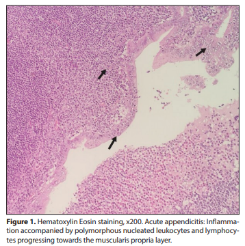
All appendectomy materials were examined histologically in our hospital within a routine procedure: The specimens were fixated in 10% formalin for 24 hours and four samples of five microns thickness were dissected for evaluation. After macroscopic evaluation, Hematoxylin-eosin stained slides were examined under a microscope. Clinical diagnoses and microscopic examination results were recorded. The cases in which neutrophil infiltration was observed in the muscularis propria of the appendix were defined as “AA” histopathologically (Figure 1). The specimens of the two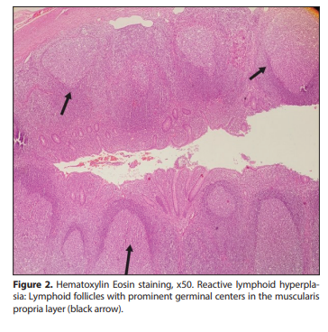
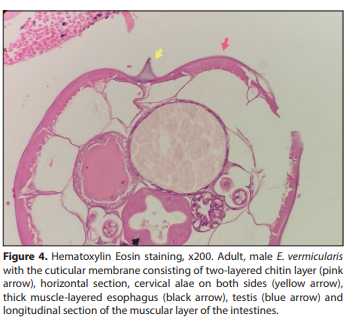
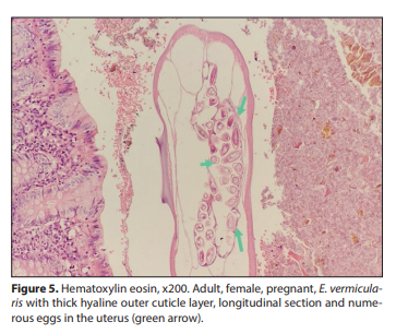
groups were re-evaluated and reported as LH (Figure 2), congestion, periappendicitis, hemorrhagic appendicitis, perforated appendicitis, abscess, phlegmonous appendicitis, gangrenous appendicitis, necrotizing appendicitis and E. vermicularis with or without peritonitis (Figure 3). The samples having hemorrhage, perforation, abscess, necrotizing, phlegmanous and/or gangrenous features were defined to be “complicated appendicitis.” E. vermicularis cases underwent additional procedures: Histochemical staining was performed with Periodic acid- Schiff (PAS) dye to evaluate the cuticular membrane consisting of a two-layered chitin layer, thick musclelayered esophagus, cervical alae, intestinal walls, uterus and testicular tissue of the nematode, which were pathognomonic in histopathological diagnosis of enterobiasis (Figure 4, 5).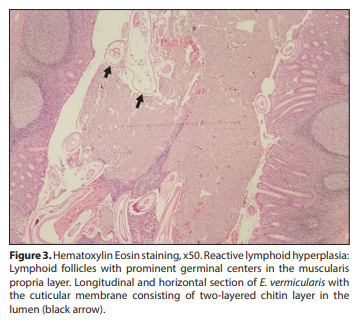
Statistical Analysis The analyses were performed by statistical package program software. The data were presented as percentages, frequencies, medians and minimum-maximum ranges or mean and standard deviation (SD) by descriptive statistics. Independent samples t-test was applied for the analyses of normally distributed continuous variables; whereas Mann-Whitney U or Kruskal Wallis tests were applied for the evaluation of abnormally distributed variables with Bonferroni correction when indicated. Cross-tables with chi-square test (χ2) and Fisher’s exact test were performed for the evaluation of categorical variables. Probability factor (p) less than 0.05 was regarded to be statistically significant. The study was approved by the Ethics Committee of NonInvasive Clinical Research of Amasya University (registration no: E-76988455-050.01.04-135458).
RESULTS
Mean age of the whole study population was 10.15 ± 3.54 years. The sex ratio of the study and control groups was man/ female: 0.84/ 0.70. The difference in sex was not statistically significant (p= 0.84). Most of the study group members lived in rural areas (n= 33; 71.7%), and 31 (67.4%) members of the control group lived in urban areas, which was statistically significant (p< 0.0001). The rate of high-income level was higher in the control group (p= 0.001). Most of the population in both groups had crowded families (number of household members ≥6). The frequency of children having their own room was significantly higher in the control group (p< 0.0001). Statistical difference was found in the water supply item (p= 0.026); the source of water supply was mains water in 84.8% (n= 39) of the control group whereas 34.8%; (n= 16) of the study group had spring water. The frequency of enterobiasis decreased significantly while the education levels of the parents increased (p< 0.0001). The data on symptoms before appendectomy were obtained from the parental on call surveys. Abdominal pain and nausea were the most common symptoms in both groups without a statistical difference (p= 0.39, p= 0.38; respectively). Pruritus ani presence was significantly higher in the study group (p< 0.0001). Vomiting, difficulty in defecation and fever were more frequent in the control group (p< 0.0001; p< 0.0001; p< 0.0001, respectively). Prevalence of growth retardation (Body mass index< 5%) was significantly higher in the study group [39.1%; (n= 18) vs. 10.9%; (n= 5); p< 0.0001]. Approximately half of the patients (n= 50; 53.4%) had radiological evaluation while the rest went through surgery on clinical diagnosis. The rate of AA was higher in the study group (32.6%; n= 15 vs. 19.6%; n= 9), which was statistically significant (p= 0.001). In addition, only one patient (2.2%) was diagnosed as “normal” in the control group; but the report of 10.9% (n= 5) of the study group was “normal”, which was a significant result (p< 0.0001). The appendiceal diameter was higher and statistically confirmed in the control group (9.05 ± 2.60 vs. 6.50 ± 1.12; p= 0.002). The average of WBC and neutrophils in CBC were higher in the control group [14530 ± 5900/mm3 ; 10500/mm3 (1500- 29300), respectively]. In addition, median of CRP levels was 1.67 mg/dL (0.5-89.9) in the study group and 12.5 mg/dL (0.25-172.8) in the control group. Elevation of acute phase reactants (WBC, neutrophils, CRP) was significantly lower in the enterobiasis group (p= 0.002; p= 0.005; p< 0.0001, respectively) whereas the median of eosinophils was higher (100/mm3 vs. 50/mm3 ; p= 0.004). There was no statistical significance in serum BUN/Cr, bilirubin, sodium and urine density levels between the groups. LH, which is thought to be reactive, was the most frequent histopathological diagnosis in the appendectomy specimens containing E. vermicularis (n= 27; 58.7%). The rate of complicated appendicitis was 73.9% (n= 34) in the control group and LH was not detected. The rates of LH, AA and complicated appendicitis were different statistically between the groups (p< 0.0001). Socio-demographic, laboratory, radiologic and histopathological features of the groups are summarized in Table 1.
DISCUSSION
E. vermicularis is a nematode requiring a human host and completing its life cycle usually in the different parts of the gastrointestinal tract. During its migration, when it visits the appendix, it may cause “appendiceal colic” and the story ends with appendectomy.
| Study Group (Enterobiasis) | Control Group (Non-enterobiasis) | p | |
|---|---|---|---|
| Sex |
Male/Female (n= 21; 45.70 / n= 25; 54.3%) |
Male/Female (n= 19; 41.3% / n= 27; 58.7%) |
0.84 |
| Number of siblings | 2 (1-5) | 2 (1-4) | 0.55 |
|
Residence
Urban area
Rural area |
19.6% (n= 9) 71.7% (n= 33) |
67.4% (n= 31) 32.6% (n= 15) |
<0.0001 |
|
Income level
Low
Intermediate High |
26% (n= 12) 58.7% (n= 27) 4.3% (n= 2) |
17.4% (n= 8) 50.0% (n= 23) 32.6% (n= 15) |
0.001 |
|
Household members
<6
≥6 |
19.6% (n= 9) 80.4% (n= 37) |
26.1% (n= 12) 73.9% (n= 34) |
0.310 |
|
Water supply
Mains water
Spring water |
65.2% (n= 30) 34.8% (n= 16) |
84.8% (n= 39) 15.2% (n= 7) |
0.026 |
|
A private room for children
Yes
No |
36.1% (n= 22) 63.9% (n= 24) |
84.8% (n= 39) 15.2% (n= 7) |
<0.0001 |
|
Maternal education
Primary education
Secondary education High education |
45.7% (n= 21) 43.5% (n= 20) 10.9% (n= 5) |
13% (n= 6) 41.3% (n= 19) 45.7% (n= 21) |
<0.0001 |
|
Paternal education
Primary education
Secondary education Higher education |
50.0% (n= 23) 37.0% (n= 17) 13.0% (n= 6) |
13% (n= 6) 39.1% (n= 18) 47.8% (n= 22) |
<0.0001 |
|
Body mass index
<5%
5-95% >95% |
39.1% (n= 18) 52.2% (n= 24) 8.7% (n= 4) |
10.9% (n= 5) 52.2% (n= 24) 37% (n= 17) |
<0.0001 |
|
Pruritus ani
Yes
No |
78.3% (n= 36) 21.7% (n= 10) |
10.9% (n= 5) 89.1% (n= 41) |
<0.0001 |
|
Abdominal pain
Yes
No |
80.4% (n= 37) 19.6% (n= 9) |
84.8% (n= 39) 15.2% (n= 7) |
0.39 |
|
Nausea
Yes
No |
82.6% (n= 38) 17.4% (n= 8) |
87.0% (n= 40) 13.0% (n= 6) |
0.38 |
|
Vomiting
Yes
No |
21.7% (n= 10) 78.3% (n= 36) |
80.4% (n= 37) 19.6% (n= 9) |
<0.0001 |
|
Difficulty in defecation
Yes
No |
21.7% (n= 10) 78.3% (n= 36) |
89.1% (n= 41) 10.9% (n= 5) |
<0.0001 |
|
Fever (≥38 °C)
Yes
No |
17.4% (n= 8) 77.6% (n= 38) |
76.1% (n= 35) 22.4% (n= 11) |
<0.0001 |
| WBC (/mm³) (mean ± SD) | 11240 ± 4030 | 14530 ± 5900 | 0.002 |
| Neutrophils (min-max) | 8590 (1790-17840) | 10500 (1500-29300) | 0.005 |
| Lymphocytes (/mm³) | 2100 ± 980 | 2200 ± 1110 | 0.655 |
| Eosinophils (/mm³) | 100 (0-800) | 50 (0-410) | 0.004 |
| Neutrophils (%) | 71.3 (4.67-94.5) | 78.1 (32.8-92.3) | 0.256 |
| Lymphocytes (%) | 20 (5-50) | 13.1 (4-55.5) | 0.064 |
| Eosinophils (%) | 1 (0-9.2) | 0.15 (0-3.6) | <0.0001 |
| CRP (mg/dL) | 1.67 (0.5-89.9) | 12.5 (0.25-172.8) | <0.0001 |
| Bilirubin (mg/dL) | 0.38 (0.1-2.1) | 0.15 (0.15-1.6) | 0.30 |
| BUN/Creatinine | 40.24 ± 20.91 | 48.04 ± 17.69 | 0.06 |
| Sodium (mEq/mL) | 137 (121-144) | 137 (131-145) | 0.584 |
| Urine density | 1020 (1010-1030) | 1018 (1001-1065) | 0.819 |
| Ultrasonographic appendix diameter | 6.5 ± 1.12 | 9.05 ± 2.60 | 0.002 |
|
Ultrasonographic diagnosis
AA
Normal |
19.6%; n= 9 10.9%; n= 5 |
32.6%; n= 15 2.2%; n= 1 |
0.001 <0.0001 |
|
Histopathological evaluation
AA
Complicated appendicitis Reactive lymphoid hyperplasia |
37.9%; n= 17 4.3%; n= 2 58.7%; n= 27 |
26.1%; n= 12 73.9%; n= 34 0%; n= 0 |
<0.0001 |