
 TR
TR
 EN
EN


ABSTRACT
Objective
Tuberculasis peritonitis is a rare clinical entity in children. There are still difficulties in diagnosis. Therefore, pediatric cases diagnosed with tuberculasis peritonitis by laparoscopic biopsy were evaluated in this study.
Materials and Methods
Five pediatric cases diagnosed with tuberculasis peritonitis in our clinic between 2005 and 2024 were included in the study. Data regarding patients’ demographic, clinical, laboratory findings, diagnostic tests, and post-discharge clinical follow-up were reviewed from hospital records.
Results
Three of the patients were female and two were male. Mean age was 12 ± 3 years. Median duration of complaints was 57 days (range: 15 and 90 days). No history of tuberculosis contact was detected in any case. The most common complaint was abdominal distension. Ascites was the most common finding on physical examination. Tuberculin skin test and interferon gamma release test were positive in only one case. The average adenosine deaminase level in the ascitic fluid was 65 UI/ dL. In this case, tuberculasis bacilli were isolated in ascitic fluid and sputum culture. The serum/ascites albumin gradient was below 1.1 g/dL in all cases. There was ascites on abdominal ultrasonography, and peritoneal thickening with ascites on abdominal computed tomography in all patients. Laparoscopic examination revealed peritoneal thickening and tuberous structures. Peritoneal biopsies revealed caseating chronic granulomatous inflammatory lesions consistent with a diagnosis of peritoneal tuberculosis. Median time to diagnosis was 14 days.
Conclusion
Tuberculasis peritonitis should also be considered in the differential diagnosis in patients with ascites. Early diagnosis and treatment are possible thanks to laparoscopic biopsy together with laboratory and radiological examinations. Mortality and morbidity related to the disease can be prevented in this way.
KEYWORDS
Tuberculasis peritonitis, child, laparoscopy, treatment
INTRODUCTION
Tuberculosis (TB) is an infectious disease as old as humanity (1). TB continues to be one of the most important causes of death from infectious diseases. The World Health Organization (WHO) reported that approximately 10 million people were diagnosed with TB in 2021, and 11% of these cases were children (2). Approximately 1 to 2 million people die from TB annually. TB primarily affects the lungs, with 16% being extrapulmonary TB (3). Abdominal TB is seen in 6-38% of untreated pulmonary TB cases and can frequently involve the gastrointestinal system, peritoneum and mesenteric lymph nodes (4,5). Abdominal TB most commonly affects the ileocecal region, but peritoneal involvement is also seen in 50- 83% of patients (6,7). TB peritonitis is rare in children. Almost all patients with TB peritonitis have clinical or subclinical ascites that develop slowly and progressively, and the most common complaint at presentation is abdominal distension (6). The most common finding on physical examination is ascites. Ascites has been reported in 93% of cases, even in all with peritoneal TB (8). In this study, five pediatric cases who presented with abdominal distension and were diagnosed with TB peritonitis by laparoscopic biopsy are presented.
MATERIALS AND METHODS
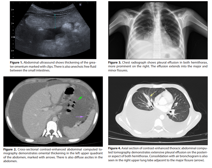
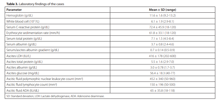
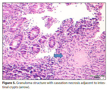
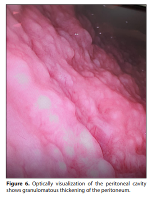
Five cases diagnosed with TB peritonitis were examined. Three of the cases were female and two were male, and median age was 12 years (range: 7.5 to 16 years). All cases presented with complaints of abdominal distension. In addition, three cases had complaints of abdominal pain and weight loss. One case had respiratory distress. Two of the cases had cough, night sweats, and fever. Median duration of symptoms was 57 days (range: 15 to 90 days). Median time to diagnosis was 14 days (range: 12 to 15 days). None of the cases had a history of contact with TB. Demographic data and clinical findings of the cases are summarized in Table 1. All cases had TST, but only two of the patients had a Bacillus Calmette-Guérin vaccination scar at the time of presentation. Induration >15 mm was observed in only one case; this case was using adalimumab (third generation tumor necrosis factor inhibitor) for uveitis due to juvenile rheumatoid arthritis. Abdominal ultrasonography revealed diffuse ascites in the abdomen in all cases (Figure 1). Abdominal computed tomography revealed heterogeneity, thickening and diffuse ascites in the omentum in all cases (Figure 2). Bilateral extensive pleural effusion due to lung involvement was detected in one case (Figure 3). Thoracic computed tomography of the patient revealed extensive pleural effusion in both hemithorax and air bronchogram adjacent to the major fissure in the upper lobe of the right lung (Figure 4). Bilateral chest tube was inserted to drain the pleural effusion in this case. Radiological findings of the cases are presented in Table 2. Paracentesis was performed in all cases. In all cases, ascitic fluid was observed to be yellow and cloudy (exudate). Serumascitic albumin gradient (SAAG) was detected as <1.1 gr/dL. Complete blood count, blood biochemistry and ascitic fluid laboratory findings of the cases are shown in Table 3. Median adenosine deaminase (ADA) level of ascitic fluids was 65 U/L (range: 18 to 118 U/L). Microscopic examination of ascitic fluids
| Case No | 1 | 2 | 3 | 4 | 5 |
|---|---|---|---|---|---|
| Age (years) | 12 | 12 | 7.5 | 16 | 12 |
| Sex | Girl | Girl | Boy | Boy | Girl |
| Duration of complaints (days) | 60 | 30 | 15 | 90 | 60 |
| Complaints | |||||
| Abdominal distension | |||||
| Weight loss | Yes | Yes | No | No | Yes |
| Abdominal pain | Yes | Yes | No | No | Yes |
| Cough | Yes | Yes | No | No | No |
| Night sweats | Yes | Yes | No | No | No |
| Respiratory distress | Yes | No | No | No | No |
| Fever | Yes | Yes | No | No | No |
| BCG scar | Yes | No | No | No | Yes |
| Tuberculin PPD test results | Negative | Positive (15 mm) | Negative | Negative | Negative |
| Time of diagnosis (days) | 15th | 12nd | 13rd | 15th | 15th |
| Case No | 1 | 2 | 3 | 4 | 5 |
|---|---|---|---|---|---|
| Chest radiograph | Normal | Pleural effusion in both hemithoraxes, prominent on the right | Normal | Normal | Normal |
| Abdominal ultrasonography | Diffuse free fluid in the peritoneal cavity | Diffuse free fluid in the peritoneal cavity | Diffuse free fluid in the peritoneal cavity | Diffuse free fluid in the peritoneal cavity | Diffuse free fluid in the peritoneal cavity and thickening of the greater omentum |
| Contrast-enhanced abdominal tomography | Thickening of the omentum and diffuse free fluid in the peritoneal cavity | Thickening of the omentum and diffuse free fluid in the peritoneal cavity | Thickening of the omentum and diffuse free fluid in the peritoneal cavity | Thickening of the omentum and diffuse free fluid in the peritoneal cavity | Thickening of the omentum and diffuse free fluid in the peritoneal cavity |
| Thoracic tomography | Cavitary lesion in the lungs | Extensive pleural effusion in both hemithoraxes and air bronchogram adjacent to the major fissure in the upper lobe of the right lung | Normal | Normal | Normal |
| Laparoscopic findings | Inflammation in the peritoneum and serosal membranes | Diffuse increased fragility of the peritoneum; adhesions and granulomatous thickening between intestinal loops and peritoneum | Diffuse increased fragility of the peritoneum; adhesions and granulomatous thickening between intestinal loops and peritoneum | Granulomatous thickening of the intestinal loops and omentum | Diffuse increased fragility and granulomatous thickening of the peritoneum |
| Peritoneal pathological examination | Non-necrotizing granulomatous peritonitis and sparsely located bacilli in AFB stain | Non-necrotizing granulomatous peritonitis | Non-necrotizing granulomatous peritonitis | Non-necrotizing granulomatous peritonitis | Non-necrotizing granulomatous peritonitis |
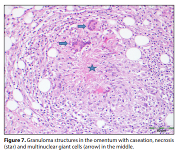
revealed abundant lymphocytes in three cases and abundant polymorphnuclear leukocytes in two cases. TB bacilli grew in the ascitic fluid and sputum cultures of one case. In this case, sparsely located bacilli in acid-fast bacilli (AFB) stain were seen in the peritoneal biopsy. TB bacilli polymerase chain reaction (PCR) was negative in ascitic fluid in all cases. A patient receiving adalimumab for uveitis due to juvenile rheumatoid arthritis had abdominal pain and diarrhea. This patient underwent colonoscopy after bowel preparation. Colonoscopy showed an edematous and hyperemic ileocecal valve. The terminal ileum could not be entered because of the risk of perforation. Histopathological examination of colon biopsies showed necrotizing granulomatous inflammation (intestinal TB) (Figure 5). Colonoscopy performed on this patient after treatment for TB showed complete resolution of the old lesions. All patients underwent diagnostic laparoscopy. During laparoscopic examination, it was observed that there was free fluid in the abdomen and the peritoneum was thick and fragile. Visualization with optically inserted trocar revealed adhesions and granulomatous thickening between the intestinal rings and the peritoneum in the abdomen that did not allow imaging (Figure 6). Histopathological examination of peritoneal biopsies revealed non-necrotizing granulomatous peritonitis (Figure 7). All cases received quadruple anti-TB treatment for 12 months. Urticarial reactions were observed in two cases due to isoniazid. In these two cases, isoniazid was stopped for a certain period and then restarted. One case with lung involvement was given corticosteroid treatment for four weeks. The cases were observed to recover completely with treatment. No problems were observed in the one-year follow-up after completion of treatment.
RESULTS
Five cases diagnosed with TB peritonitis were examined. Three of the cases were female and two were male, and median age was 12 years (range: 7.5 to 16 years). All cases presented with complaints of abdominal distension. In addition, three cases had complaints of abdominal pain and weight loss. One case had respiratory distress. Two of the cases had cough, night sweats, and fever. Median duration of symptoms was 57 days (range: 15 to 90 days). Median time to diagnosis was 14 days (range: 12 to 15 days). None of the cases had a history of contact with TB. Demographic data and clinical findings of the cases are summarized in Table 1. All cases had TST, but only two of the patients had a Bacillus Calmette-Guérin vaccination scar at the time of presentation. Induration >15 mm was observed in only one case; this case was using adalimumab (third generation tumor necrosis factor inhibitor) for uveitis due to juvenile rheumatoid arthritis. Abdominal ultrasonography revealed diffuse ascites in the abdomen in all cases (Figure 1). Abdominal computed tomography revealed heterogeneity, thickening and diffuse ascites in the omentum in all cases (Figure 2). Bilateral extensive pleural effusion due to lung involvement was detected in one case (Figure 3). Thoracic computed tomography of the patient revealed extensive pleural effusion in both hemithorax and air bronchogram adjacent to the major fissure in the upper lobe of the right lung (Figure 4). Bilateral chest tube was inserted to drain the pleural effusion in this case. Radiological findings of the cases are presented in Table 2. Paracentesis was performed in all cases. In all cases, ascitic fluid was observed to be yellow and cloudy (exudate). Serumascitic albumin gradient (SAAG) was detected as <1.1 gr/dL. Complete blood count, blood biochemistry and ascitic fluid laboratory findings of the cases are shown in Table 3. Median adenosine deaminase (ADA) level of ascitic fluids was 65 U/L (range: 18 to 118 U/L). Microscopic examination of ascitic fluids revealed abundant lymphocytes in three cases and abundant polymorphnuclear leukocytes in two cases. TB bacilli grew in the ascitic fluid and sputum cultures of one case. In this case, sparsely located bacilli in acid-fast bacilli (AFB) stain were seen in the peritoneal biopsy. TB bacilli polymerase chain reaction (PCR) was negative in ascitic fluid in all cases. A patient receiving adalimumab for uveitis due to juvenile rheumatoid arthritis had abdominal pain and diarrhea. This patient underwent colonoscopy after bowel preparation. Colonoscopy showed an edematous and hyperemic ileocecal valve. The terminal ileum could not be entered because of the risk of perforation. Histopathological examination of colon biopsies showed necrotizing granulomatous inflammation (intestinal TB) (Figure 5). Colonoscopy performed on this patient after treatment for TB showed complete resolution of the old lesions. All patients underwent diagnostic laparoscopy. During laparoscopic examination, it was observed that there was free fluid in the abdomen and the peritoneum was thick and fragile. Visualization with optically inserted trocar revealed adhesions and granulomatous thickening between the intestinal rings and the peritoneum in the abdomen that did not allow imaging (Figure 6). Histopathological examination of peritoneal biopsies revealed non-necrotizing granulomatous peritonitis (Figure 7). All cases received quadruple anti-TB treatment for 12 months. Urticarial reactions were observed in two cases due to isoniazid. In these two cases, isoniazid was stopped for a certain period and then restarted. One case with lung involvement was given corticosteroid treatment for four weeks. The cases were observed to recover completely with treatment. No problems were observed in the one-year follow-up after completion of treatment.
DISCUSSION
In the 2020 Türkiye Tuberculosis Control Report, it was reported that the total number of TB cases in our country was 11.786 and the incidence of TB was 14.1 per hundred thousand (10). TB peritonitis is reported in approximately 3.5% of patients with pulmonary TB and 31-58% of patients with abdominal TB. TB peritonitis can be seen in 1% of all patients with TB (5). Gürkan et al. published 11 cases of children with TB peritonitis in 1999 (11). After that, Dinler et al. reported nine cases of children with TB peritonitis from our country in 2009 (12). In the following years, pediatric cases diagnosed with TB peritonitis in our country were presented as case reports (13-17). These studies support the fact that TB peritonitis in children is rare in our country, as in the world. Peritoneal TB is usually seen in children with immune deficiency (8). In the present study, one patient was using adalimumab due to juvenile rheumatoid arthritis. Studies have reported that the ages of children followed up for TB peritonitis ranged from 9 to 14 years (11-14). Mean age of the cases in the present study was 12, which is consistent with this article. Patients with TB peritonitis most commonly present with complaints of abdominal distension, abdominal pain, and weight loss (12). In the present study, all children had abdominal distension and three had abdominal pain and weight loss. The data in the present study were consistent with the literature (8,12,17). A history of contact with a TB case was reported in 66.6% of the cases with TB peritonitis (12). However, none of our cases had a history of contact. Among all cases in which TST was performed, only one case was found to have an induration >15 mm. It was thought that the negative TST determination in the other four cases might have been due to differences in the applied technique, solution used and interpretation (18). In laboratory tests, no disease-specific findings were observed in complete blood count. White blood cell count is usually within normal range. In the present study, white blood cell count was found to be normal. Data in the present study were consistent with the literature (12). Erythrocyte sedimentation rate is usually increased (19). It was found to be significantly elevated in three of our cases. Pulmonary involvement is reported in 12-55% of cases with TB peritonitis (12,15). In the present study, pulmonary involvement (pleural effusion) was observed in only one case, consistent with the literature. The bilateral chest tube inserted in this case was removed after the pleural effusion regressed with treatment. Radiological imaging techniques such as abdominal ultrasonography and computed tomography provide very useful information in the diagnosis of TB peritonitis. The most common finding on abdominal ultrasonography is free fluid accumulation in the abdomen (20). All patients in the present study had ascites in their abdomen on abdominal ultrasonography. Abdominal computed tomography shows free fluid, omental thickening, and enlarged lymph nodes in the abdomen due to TB peritonitis (20-22). In this study, radiological findings were consistent with the literature. Paracentesis was performed in all cases. The ascitic fluid was found to be exudative, rich in lymphocytes, and SAAG was below 1.1 g/dL. The data obtained in present study were consistent with the literature (23). It is thought that these findings may be reliable parameters in the diagnosis of TB peritonitis. The most reliable method for the diagnosis of TB is the culture of TB bacillus (Mycobacterium tuberculosis) from body fluids. On direct microscopic examination of ascitic fluid, AFB can be seen in less than 2% of cases (24). It is reported that TB bacillus growth in culture is between 0-83% (25). In our study, AFB was detected in the ascitic fluid of one case and M. tuberculosis grew in the ascitic fluid of the same case. In noncirrhotic cases, measurement of ADA in ascitic fluid remains a highly reliable laboratory test for the diagnosis of TB peritonitis (26). In the present study, ADA levels were found to be elevated in 80% of cases, consistent with the literature. In cases of TB peritonitis, low AFB positivity in ascitic fluid leads to a decrease in the sensitivity of the PCR test (27). In the present study, AFB PCR positivity was not detected in any case of ascitic fluid. Laparoscopic examination is the most commonly used method for peritoneal imaging and peritoneal biopsy (28). It continues to be the most appropriate method for early and definitive diagnosis of the disease (12). The most common findings in laparoscopic examination are ascites, peritoneal thickening, adhesions, and millimetric tuberous structures (27). In the present study, laparoscopic examination findings of our cases were consistent with the literature. Pathological examination of chronic granulomatous inflammatory reaction with caseation is highly specific for TB peritonitis (29). Similar findings were observed in the pathological examination of peritoneal biopsy in our cases. It has been reported that corticosteroid treatment reduces complications and mortality in cases of TB peritonitis (26). In the present study, it was observed that four weeks of corticosteroid treatment accelerated recovery in a case with bilateral pleural effusion. TB continues to be an important health problem worldwide, especially in underdeveloped and developing countries. Although TB peritonitis is not common in children, it is a disease that should be considered in the differential diagnosis of children presenting with abdominal distension due to ascites. Mortality and morbidity can be prevented through early diagnosis and treatment with laparoscopic examination in addition to history, physical examination and laboratory tests.