
 TR
TR
 EN
EN


ABSTRACT
Objective
This study aimed to investigate the clinical characteristics of pediatric patients diagnosed with acute respiratory infections in the Pediatric Intensive Care Unit at Bakırköy Dr. Sadi Konuk Training and Research Hospital, whose nasopharyngeal swab samples tested positive for human bocavirus (HBoV). The prevalence of HBoV as a solitary infection or in co-infection and its clinical distinctions were examined.
Materials and Methods
The study included 50 children who were monitored in the Pediatric Intensive Care Unit of Bakırköy Dr. Sadi Konuk Training and Research Hospital between 2015 and 2021 diagnosed with acute respiratory infections and were found to be positive for HBoV in nasopharyngeal swab samples. Demographic and clinical information for all patients was collected, and symptoms such as respiratory distress, cough, and fever were recorded. Nasopharyngeal swab samples were analyzed using a polymerase chain reaction panel that tested for 14 different viruses to determine the presence of HBoV and other viral agents. Patients with solitary HBoV infection and those with detected coinfections were compared to assess clinical progressions and laboratory findings. Statistical analyses were conducted using IBM SPSS Statistics 22 (IBM SPSS, Türkiye), with a significance level set at p< 0.05.
Results
It was determined that 86% of the 50 patients included in the study were under the age of three. Rhinovirus was identified as the most common co-infecting agent. Patients in the co-infection group exhibited significantly longer durations of symptoms (p= 0.014), and the frequency of atelectasis was notably higher in the co-infection group (p= 0.009). In the group with sole HBoV infection, eosinophil levels were found to be significantly higher (p= 0.002).
Conclusion
This study demonstrates that HBoV can appear both as a sole infection and in co-infection in children suffering from acute respiratory infections. No significant differences in clinical symptoms were observed between the groups. The duration of breastfeeding was found to be longer in the group with sole HBoV infection. Patients in the co-infection group exhibited longer durations of symptoms. Patients with only HBoV infection had a higher absolute eosinophil count while a higher incidence of atelectasis was observed in the co-infection group. These findings suggest the need for further studies to confirm these observations.
KEYWORDS
Human bocavirus, paediatric intensive care, acute respiratory tract infection
INTRODUCTION
Acute respiratory infections represent a significant source of mortality and morbidity among children globally. Viruses are known to be responsible for approximately 80% of all acute respiratory infections. The most frequently identified viruses are rhinovirus, respiratory syncytial virus (RSV), influenza, parainfluenza, metapneumovirus, and adenovirus. However, recent advances in polymerase chain reaction (PCR) research have facilitated the identification of new viruses (1). Human bocavirus (HBoV), first identified in 2005 among children with acute respiratory infections, is categorized as a non-enveloped, single-stranded DNA virus within the Parvoviridae family (1). Of the HBoV types, HBoV-1 is associated with respiratory infections, while HBoV-2, HBoV-3, and HBoV-4 have been found in fecal samples and are recognized as causes of gastroenteritis. From 2005 to 2024, HBoV was detected in 3.1% to 22.5% of nasopharyngeal aspirate samples from children diagnosed with acute respiratory infections (1,2). The virus predominantly affects children under five years old, especially those with lower respiratory tract infections, and is implicated in the pathogenesis of severe clinical conditions such as bronchiolitis, pneumonia, and asthma exacerbations. While it can be detected year-round, incidences peak during the winter and spring months (3). There are studies suggesting that respiratory infections caused by HBoV can be as severe as those caused by other viruses, though others indicate no significant difference in clinical severity (4). Research has frequently demonstrated that HBoV is commonly detected in respiratory samples alongside other viruses. A prospective study published in 2010 found HBoV in 33% of 318 acute respiratory disease cases, with 72% of these cases also testing positive for other viruses. The detection of HBoV in 44% of samples from asymptomatic individuals in this study has led to the hypothesis that HBoV might be more of a colonizer than a pathogen, influencing subsequent research directions (5,6). Later studies have identified a strong correlation between HBoV viral load and disease severity, establishing it as a respiratory pathogen (7). Furthermore, another study conducted in 2010 investigated HBoV serology in patients suffering from respiratory infections and demonstrated an increase in immunoglobulin G titers, indicating HBoV’s role as a causative agent in respiratory infections (8). Research has shown that HBoV can be detected in respiratory samples for up to 75 days in patients monitored on an outpatient basis and for up to 4.5 months in those hospitalized (5). The frequent detection of HBoV along with other viruses in symptomatic patients, and its presence in asymptomatic individuals, may be explained by this prolonged shedding. Research supports the idea that the detection of HBoV alone, the demonstration of viremia, and a high viral load could be indicative of an actual HBoV infection (9,10). However, more comprehensive research is necessary to better understand the clinical progression and characteristics of this virus and its co-infections. In this study, we examined patients who were monitored in our hospital’s pediatric intensive care unit for acute respiratory infections between 2015 and 2021 and tested positive for HBoV in nasopharyngeal swab samples. Our aim was to describe the clinical and serological features of these patients and to investigate the differences between cases of HBoV as a sole infection and as a co-infection.
MATERIALS AND METHODS
Our study was designed as a single-center, retrospective analysis. Between 2015 and 2021, 50 pediatric patients diagnosed with acute respiratory infections and admitted to the pediatric intensive care unit of hospital, who tested positive for HBoV in nasopharyngeal swab samples, were included in the study. Information such as age, birth weight, gestational age, known medical history, medication use, any visits to other healthcare facilities within the last week, prior nebulizertreatments, and family history of asthma or atopy was collected from the families and recorded. Upon admission to the intensive care unit, PRISM scores, blood gas analyses, complete blood counts, C-reactive protein levels, immunoglobulin levels, and chest radiography findings of the patients were also documented. Throughout their clinical course, the patients’ needs for non-invasive and invasive mechanical ventilation, as well as total length of stay, were monitored and recorded. All nasopharyngeal swab samples from the patients were analyzed at the Microbiology Laboratory of Hospital using a real-time (RT) PCR panel that covers 14 viruses, including SARS-CoV, influenza virus, parainfluenza virus, rhinovirus, enterovirus, adenovirus, human metapneumovirus, and HBoV. The nasopharyngeal swab sample was tested using the Bioeksen RT-qPCR kit. It should be stated that informed consent was obtained from the legal guardians of all patients included in the study. The patients included in the study were divided into two groups: those with only HBoV detected and those with HBoV plus other viruses detected, and the data were compared between these groups. Statistical analyses were conducted using IBM SPSS Statistics 22 (IBM SPSS, Türkiye). Median, interquartile ranges, and range between quartiles were calculated for all continuous variables. The normality of data distribution was assessed using the Shapiro-Wilk test. Descriptive statistical methods (mean, standard deviation, frequency) were utilized to summarize the data; quantitative data comparisons between the two groups for normally distributed parameters were performed using the Student’s t-test, and for non-normally distributed parameters using the Mann-Whitney U test. The chisquare test was used for the comparison of qualitative data. The level of statistical significance was set at p< 0.05.
RESULTS
Fifty patients admitted to the pediatric intensive care unit with a diagnosis of acute respiratory tract infection between 2015 and 2021 were included in the study, all of whom tested positive for HBoV in nasopharyngeal swab samples. Mean age of the patients was 13.9 months, with the majority being under three years of age. Most presented within two days of symptom onset, and respiratory distress was the predominant clinical finding (Table 1). Upon evaluating the timing of patient admissions, 70% were found to occur during the winter and autumn seasons. There was no statistically significant difference in the distribution of admission months between patients with isolated HBoV infection and those with co-infection (p= 0.56) (Table 2). PCR analysis of nasopharyngeal swab samples collected from all patients revealed that HBoV was identified as the sole 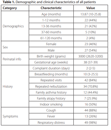

pathogenic agent in 56% of cases. Rhinovirus was identified as the most frequently co-detected virus (Figure 1). Single HBoV was the most frequently detected virus, observed in 56% of cases. Rhinovirus was identified in 20% of the patients while adenovirus accounted for 6%. Other viruses, including coronavirus, enterovirus, metapneumovirus, and parainfluenza, were each detected in 4%, and influenza was found in 2%. Patients with single HBoV infection had a mean age of 15.8 months while those with co-infections had a mean age of 13.7 months, with no significant age or sex differences between the groups. The duration of symptoms was significantly longer in patients with co-infections (p= 0.014). In contrast, the duration of breastfeeding was significantly longer in the single-infection group (p= 0.047). Other demographic and clinical features were comparable between the groups (Table 3).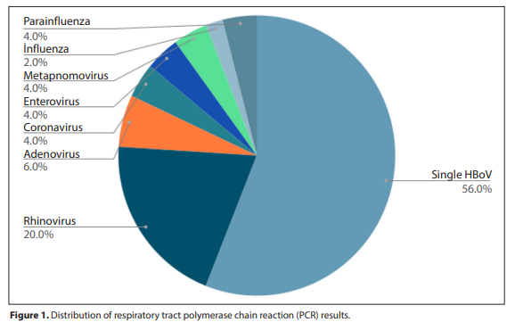
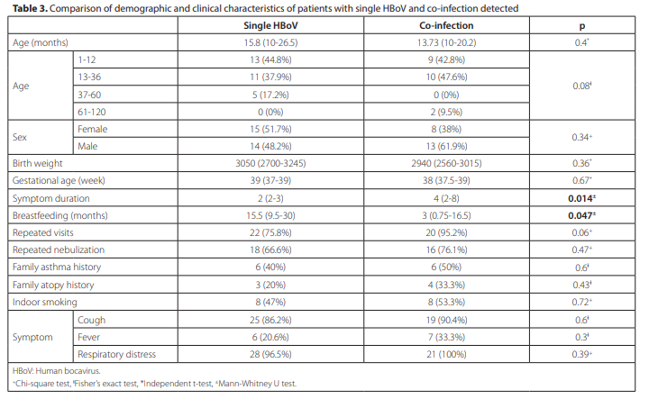
No significant differences were observed between the patients with single HBoV infection and those with co-infections in terms of PRISM scores, vital signs, blood gas results, acute phase reactants, hematological or immunological parameters (p> 0.05). The eosinophil count was significantly higher in the single infection group (p= 0.002), and eosinophilia (≥300 cells/μL) was more frequent in this group, although not statistically significant (p= 0.06). Radiologically, atelectasis was 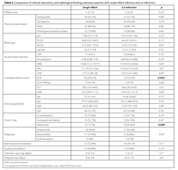
significantly more common among patients with co-infections (p= 0.009). Any additional viral pathogen identified by PCR was defined as a coinfection. No significant differences were found between the groups regarding the need for respiratory support or the length of intensive care and hospital stay (p> 0.05) (Table 4).
DISCUSSION
Our study demonstrated that HBoV can be detected alone or in conjunction with other viruses in respiratory tract PCR samples of children admitted to the pediatric intensive care unit with a diagnosis of acute respiratory infection. The average age of the patients was determined to be 13.8 months, with 86% under the age of three and 54% being male. In a study conducted in Barcelona in 2023, similar to our study, 60% of patients with detected HBoV were male, and 88% were under the age of three (11). Another study in 2013 found that the average age of the patients was 7.9 months, with 55% being male (4). Our findings are consistent with the literature in this respect.In our study, 70% of our patients required repeated nebulization treatments, and 50% were exposed to household smoking. Similar results were shown in another study conducted in our country in 2023. When examining patients who were hospitalized due to respiratory infections associated with HBoV, 70% had accompanying illnesses, and 50% of these patients required hospitalization due to severe pneumonia (12). Another study involving hospitalized patients found that 17% had a history of repeated nebulization treatments, and these patients significantly more often required intensive care admissions (13). Since the patients included in our study were monitored in pediatric intensive care, a higher incidence of episodic wheezing history was considered an expected outcome. It has been shown that HBoV infections are common during the winter and autumn months. Consistent with the literature, the majority of patients in our study also presented during the winter and autumn months. When examining seasonal distribution between the single infection and co-infection groups, no statistically significant difference was found, aligning with similar studies (1,10,12). In our study, at least one virus was detected accompanying HBoV in 44% of the patients. The most frequently accompanying virus was Rhinovirus. Adenovirus, coronavirus, enterovirus, metapneumovirus, parainfluenza, and influenza virus were other viruses detected at similar rates. In the literature, the co-infection rate varies between 41.3% and 95% (4,13,14). In a large cohort study conducted in Saudi Arabia, co-infections were detected in over 80% of the patients. Similar to our findings, rhinovirus was the most common co-pathogen, followed by adenovirus (15). Another similar study revealed that Rhinovirus and RSV were the most frequent co-infecting agents (16). When comparing patients with single HBoV infection to those with co-infections, no significant differences were found in age and sex between the two groups, consistent with the literature. However, the duration of symptoms was significantly shorter in the single HBoV infection group (p= 0.014). This suggests that patients with single HBoV infections required admission to the pediatric intensive care unit shortly after symptom onset, indicating a potentially faster progression compared to co-infections. Although previous studies have shown that co-infections tend to have a quicker and more severe clinical course, our study does not support this aspect of the literature (17). Our study demonstrated that patients with co-infections received breast milk for a significantly shorter duration (p= 0.047). A review of the literature did not yield similar findings. However, it is well-known that breast milk contains immunological components that provide protection against respiratory infections, reducing the duration and severity of infections. It is therefore expected that prolonged breastfeeding could offer protection against multiple infections. In our study, the severity of patients’ clinical conditions upon admission to the unit was assessed using PRISM scoring. Although the mortality risk was higher in the co-infection group, no statistically significant difference was found between the two groups. When comparing the need for mechanical ventilation, as well as the duration of intensive care and hospital stays between the two groups, no statistically significant differences were identified. A cohort study published in 2024 involving 165 patients demonstrated that co-infections were associated with longer mechanical ventilation and extended hospital stays (13). The inability to replicate similar results in our study may be related to an insufficient sample size. Physical examination findings indicated that rales were more prominent in the single HBoV group, while extended expiration was more frequently observed in the co-infection group, though these findings were not statistically significant. In both groups, pneumonia was the most common diagnosis. Consistent with the literature, HBoV is most frequently associated with upper respiratory tract infections, and in cases of lower respiratory tract infections, it is most commonly linked to pneumonia and bronchiolitis (12,13). In our study, when comparing laboratory tests of the patients, no statistically significant differences were observed except for the absolute eosinophil count. The absolute eosinophil count was notably higher in the single HBoV infection group (p= 0.02). However, when comparing patients with an absolute eosinophil count above 300/μL, no statistically significant difference was found (p= 0.06). A study published in 2020 compared single HBoV infections with RSV-HBoV co-infections and, although no significant difference in eosinophil counts was detected between the two groups, the counts were slightly higher in the single HBoV group (18). When comparing chest X-rays of the patients, atelectasis was more frequently observed in the co-infection group (p= 0.009). A study published in 2020, which compared patients with viral lower respiratory tract infections monitored with high-flow nasal cannulas, found that co-infections increased the severity of the disease and were more frequently associated with atelectasis (19). This study has certain limitations. The relatively small sample size and retrospective design should be taken into consideration. It is difficult to determine how long these viral pathogens remain PCR-positive in the nasopharynx, and it should be kept in mind that the patient’s immune response may influence this duration. Therefore, the development of PCR techniques capable of quantifying viral copy numbers and enabling PCR analysis from blood samples could provide more comprehensive and quantitative insights into the pathogenic potential of these viruses. Prospective, large-scale studies are warranted to further clarify these findings.In conclusion, our study demonstrated that HBoV can be detected in children experiencing acute respiratory infections, both as a single infection and in co-infections. No significant differences in clinical symptoms were observed between the groups. The duration of breastfeeding was found to be longer in the group with single HBoV infection. It was observed that patients in the co-infection group had longer durations of symptoms. Patients in the single HBoV infection group had a higher absolute eosinophil count while atelectasis was more frequently observed in the co-infection group. In conclusion, our study demonstrated that HBoV can be detected in children experiencing acute respiratory infections, both as a single infection and in co-infections. No significant differences in clinical symptoms were observed between the groups. The duration of breastfeeding was found to be longer in the group with single HBoV infection. It was observed that patients in the co-infection group had longer durations of symptoms. Patients in the single HBoV infection group had a higher absolute eosinophil count while atelectasis was more frequently observed in the co-infection group. Further research is needed to validate these findings.