
 TR
TR
 EN
EN


INTRODUCTION
A two-month-old male infant was brought to the pediatric outpatient clinic due to a gradually enlarging swelling in the midline of the abdomen, noted since the second week after birth. The family reported no fever, vomiting, diarrhea, or feeding difficulties. The infant had been born at term via spontaneous vaginal delivery, with an uneventful perinatal period. There was no history of umbilical venous or arterial catheterization, omphalitis, or abdominal intervention. Vital signs were normal. On physical examination, a small umbilical hernia was present; there was no erythema, edema, or discoloration of the abdominal skin, but marked tenderness was noted upon palpation in the region of the swelling. Other systemic findings were normal. Laboratory evaluation revealed leukocytosis (WBC: 20.000/mm³) with neutrophil predominance and elevated C-reactive protein (CRP) (55 mg/L); other parameters were within normal limits. On abdominal ultrasound (US), a lesion measuring 57 × 35 mm was detected in the midline and left paramedian region, extending along the course of the falciform ligament between the anterior surface of the left hepatic lobe and the anterior abdominal wall. There was a marked increase in echogenicity surrounding the lesion. Color Doppler examination demonstrated intense vascularity both peripherally and centrally along a linear component (Figure 1). Although the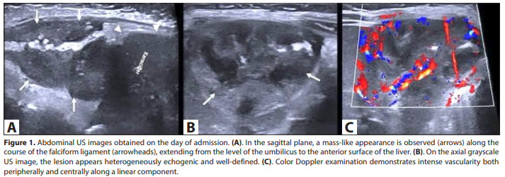
localization of the lesion along the falciform ligament and the surrounding increased echogenicity suggested an infectious process, magnetic resonance imaging (MRI) was planned for detailed evaluation and to exclude other causes originating from the falciform ligament. Five days after admission, abdominal MRI was performed under sedation. A well-defined, midline lesion was identified between the anterior surface of the liver and the anterior abdominal wall, extending along the expected course of the falciform ligament. The central portion was consistent with liquefaction. The peripheral zone demonstrated intermediate signal characteristics compatible with granulation tissue and a reactive inflammatory wall. The visualization of the ligamentum teres as a thickened, hypointense linear structure along the inferior free margin of the falciform ligament further supported ligament involvement (Figure 2). There was fat stranding and T2 signal increase in the surrounding tissues of the falciform ligament. Fluid collection anterior to the muscle planes of the abdominal wall was also present. No pathological contrast enhancement was observed in the adjacent hepatic parenchyma, and no intrahepatic abscess extension or portal vein thrombosis was detected. Based on the patient’s history, clinical findings, and radiological features, what is your diagnosis? DIAGNOSIS: Falciform ligament abscess Brief Discussion The falciform ligament is a sickle-shaped peritoneal fold extending from the anterior abdominal wall to the visceral surface of the liver. Its inferior free margin contains the ligamentum teres hepatis (obliterated umbilical vein), along with small paraumbilical vessels, fatty tissue, and occasionally aberrant bile ducts (1). This close anatomical relationship with the umbilical venous system makes the falciform ligament a potential route for infection spread, particularly in neonates and infants. Falciform ligament abscess is an extremely rare clinical entity. Most reported pediatric cases are associated with umbilical venous catheterization, omphalitis, or intra-abdominal surgery; however, sporadic cases without identifiable predisposing factors have also been described (2-4). Accurate radiological characterization is crucial for both diagnosis and treatment planning. Imaging plays a decisive role at every stage of the diagnostic process. US is the first-line modality due to its accessibility, lack of ionizing radiation, and real-time imaging capability. However, as observed in our case, early-stage infection may mimic solid neoplasms on US. In such cases, MRI, with its 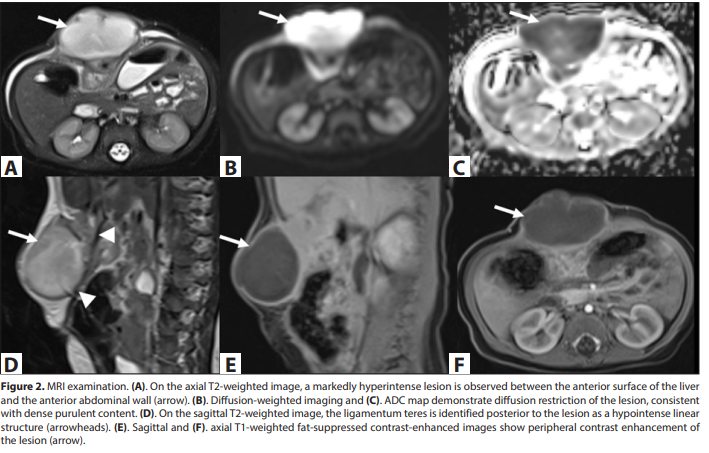
superior soft tissue contrast resolution and multiplanar imaging capability, clearly demonstrates the anatomical extent of the disease and is critical in excluding alternative diagnoses. The differential diagnosis of falciform ligament abscess includes primary tumors of the falciform ligament, hepatic abscess, incarcerated omental hernia, lymphatic malformation, and anterior abdominal wall hematoma. When US and MRI findings are evaluated together, the development of cystic features in the lesion and peripheral rim enhancement effectively exclude solid masses. Although hepatic abscess is the most important differential diagnosis due to the lesion’s proximity to the left hepatic lobe, confinement of the pathology to the falciform ligament and the normal appearance of the adjacent hepatic parenchyma on MRI ruled it out. Incarcerated omental hernia was excluded due to the absence of signal characteristics specific to omental fat, lack of hernia sac and bowel loops, and the lesion’s exact correspondence with the falciform ligament course. Anterior abdominal wall hematoma was excluded based on the absence of trauma or coagulopathy history, lack of hemorrhagic signal features on MRI, and the presence of leukocytosis and elevated CRP. Lymphatic malformations are typically multilocular and do not demonstrate diffusion restriction or inflammatory clinical features unless infected. In this case, it is noteworthy that the clinical course was insidious and slowly progressive, with the infant remaining afebrile despite a symptom duration of one month. This highlights the potential for delayed diagnosis in infants who may not exhibit classical signs of sepsis. Delayed diagnosis may lead to serious complications such as hepatic abscess, peritonitis, and septicemia; therefore, radiological evaluation is indispensable not only for diagnosis but also for early detection of complications and detailed assessment of disease spread. In our patient, minimally invasive abscess drainage was performed by the pediatric surgery team, and concurrent antibiotic therapy was initiated. Staphylococcus aureus was isolated from the abscess culture. After complete resolution of clinical and laboratory findings, excision of the falciform ligament was performed, and the infant was discharged in good health.