
 TR
 EN


ABSTRACT
Objective
Methicillin-resistant Staphylococcus aureus (MRSA) is a significant hospital-acquired infection due to its morbidity and mortality. Although successful results have been achieved with the introduction of glycopeptide antibiotics, particularly vancomycin, the decrease in vancomycin susceptibility over time has increased concerns about treatment failure. This study aims to determine the frequency of methicillin-resistant isolates, heterogeneous vancomycin-intermediate S. aureus (hVISA), and vancomycin-intermediate S. aureus (VISA) in pediatric cases using advanced microbiological methods and to evaluate the clinical significance of these phenotypes.
Materials and Methods
In this retrospective study conducted at a tertiary center, pediatric patients with MRSA detected in blood and other body fluid cultures between 2001 and 2016 were evaluated. The presence of hVISA and VISA in stored isolates from patients diagnosed with MRSA was investigated using the E-test macromethod and population analysis profile–area under the curve (PAP-AUC) methods. Patients were examined in terms of parameters such as demographic characteristics, comorbid diseases, primary focus of infection, vancomycin minimum inhibitory concentration (MIC), laboratory findings, length of hospital stay, and treatment failure. Treatment failure was defined as the persistence of fever and continued growth in cultures on the seventh day, no decrease in inflammatory markers, the presence of recurrent positive cultures, and mortality. Patients were divided into two groups, and whether there were significant differences in clinical characteristics between the MRSA and hVISA/VISA groups was evaluated.
Results
Heterogeneous VISA was detected in 15 of the 68 children included in the study (22%); VISA was not detected. Bacteremia, pneumonia, and soft tissue infections were the most common infections in the hVISA group. Vancomycin MIC values were significantly higher in the hVISA group (p< 0.01). Although hospital stay was longer in the hVISA group, it was not statistically significant. Vancomycin treatment failure was observed in 60% of hVISA cases. In the hVISA group, recurrent growth continued in nine patients, and four patients died in the first 28 days.
Conclusion
Currently, due to the frequent use of vancomycin in the treatment of MRSA infections, resistance cases have begun to be encountered. In cases where adequate response to vancomycin treatment is not obtained, hVISA/VISA screenings should be performed, as they can be considered a precursor to resistance development. Although it is an expensive and laborious test, the PAP-AUC method remains the most sensitive method for detecting hVISA/VISA strains.
KEYWORDS
Vancomycin-intermediate staphylococcus aureus, vancomycin treatment failure, glycopeptide resistance
INTRODUCTION
Staphylococcus aureus is a facultative anaerobic bacterium capable of causing a wide spectrum of clinical diseases, ranging from superficial skin and soft-tissue infections to life-threatening conditions such as bloodstream infections and meningitis (1). While the introduction of penicillin initially brought victory in treating these infections, the rapid emergence of antibiotic resistance quickly created a global health problem (2).
The first reports of methicillin-resistant S. aureus (MRSA) in the early 1960s marked a turning point in clinical practice (3). Glycopeptide antibiotics, particularly vancomycin, subsequently became the cornerstone of therapy for methicillin-resistant strains. However, decreasing susceptibility to vancomycin has gradually emerged and now poses a significant therapeutic concern (4).
Among the forms of reduced susceptibility, heterogeneously vancomycin-intermediate S. aureus (hVISA) is of particular importance. These strains appear susceptible or intermediate by routine laboratory testing yet contain subpopulations with substantially reduced susceptibility that standard methods frequently fail to detect. Such heterogeneity allows a small but clinically relevant fraction of the bacterial population to survive vancomycin exposure, thereby contributing to persistent infection. For this reason, heterogeneously vancomycin-intermediate strains are widely regarded as precursors to fully vancomycin-intermediate S. aureus (VISA) (5).
The first well-characterized isolate demonstrating this phenomenon was reported in Japan in 1997, highlighting the need for more sensitive diagnostic approaches (6). Although several microbiological techniques have been developed to detect reduced susceptibility, population analysis profiling– area under the curve (PAP-AUC) remains the most reliable and widely accepted reference method as it can reveal resistant subpopulations that are not apparent with broth microdilution or standard diffusion-based assays. Despite this, the technique’s labor-intensive nature and cost have limited its routine clinical use (7).
Evidence describing the prevalence and clinical consequences of hVISA in pediatric patients remains scarce. Children are particularly vulnerable due to age-dependent differences in immune function, pharmacokinetics, and underlying comorbidities; therefore, adult data cannot be directly extrapolated to pediatric populations (8). Given the increasing recognition of reduced vancomycin susceptibility and the paucity of pediatric data, the present study aimed to re-evaluate vancomycin susceptibility among stored isolates from children with culture-confirmed MRSA infection, to identify the presence of hVISA/VISA using both screening and reference methods, and to examine the clinical features and outcomes associated with these infections.
MATERIALS AND METHODS
Study Design and Ethics Approval
This study was approved by the Hacettepe University Faculty of Medicine Ethics Committee (Decision no: GO16/323- 05, Date: 24.05.2016). The study included children from birth to 18 years of age who had culture-confirmed MRSA infection and for whom both clinical data and stored bacterial isolates were available. The study period extended from November 2001 to November 2016.
Patient Selection and Clinical Data Collection
All pediatric patients with at least one culture positive for MRSA during the study period were screened. Patients were excluded if their stored bacterial isolates were unavailable, if the organism was determined to represent colonization or contamination rather than true infection, or if clinical records were incomplete. For each eligible patient, the following clinical variables were recorded:
• Age, sex, and underlying chronic diseases
• Type of clinical specimen (sterile vs. non-sterile body fluid)
• Serum inflammatory markers including C-reactive protein (CRP) and complete blood count parameters
• Vancomycin treatment characteristics including dosing, timing of initiation, and duration of therapy
• Vancomycin minimum inhibitory concentration (MIC) values determined by each laboratory method
• Clinical response on day seven of treatment and at completion of therapy
• Length of hospital stay, recurrence of infection, treatment failure, and mortality within 28 days
• History of hospitalization or antibiotic exposure prior to the current admission
Treatment failure was defined as;
• persistent fever (fever still exceeding 38 ºC on the 7th day of treatment)
• Sustained elevation of inflammatory markers (CRP, Interleukin-6, procalcitonin)
• Persistence of positive cultures
• Death occurring by day seven or by the end of therapy.
Mortality within 28 days following diagnosis was considered infection-related unless an alternative cause was clearly documented. Recurrence was defined as continued bacterial growth in follow-up cultures after completion of an adequate therapeutic course.
Microbiological Processing of Isolates
Stored isolates had been preserved at −80 °C in brain– heart infusion agar supplemented with glycerol. For the study, isolates were thawed and sub-cultured onto sheep blood agar and incubated at 37 °C for 24 hours to ensure viability and purity. Typical colonies were confirmed as S. aureus using catalase testing and tube coagulase testing.
Determination of Vancomycin Susceptibility
To assess vancomycin susceptibility, three methods were applied to each isolate:
• Broth microdilution method
• Standard gradient diffusion method (E-test)
• Population analysis profile with area-under-the-curve calculation (reference method)
All procedures were performed in accordance with accepted clinical microbiology guidelines(9).
Broth Microdilution
Vancomycin was prepared in two-fold serial dilutions ranging from 0.125 to 64 micrograms per milliliter in Mueller–Hinton broth. Bacterial suspensions were standardized to the 0.5 McFarland turbidity standard, diluted, and inoculated into microdilution wells. After incubation at 35°C for 24 hours, the MIC was recorded as the lowest concentration demonstrating complete inhibition of visible growth. Isolates were subsequently categorized as susceptible, intermediate, or resistant based on established interpretative criteria.
Standard Gradient Diffusion Method
Vancomycin gradient diffusion strips were applied to Mueller–Hinton agar plates inoculated with standardized bacterial suspensions. Plates were incubated for 24 hours at 35°C, and the MIC was read at the point where the elliptical zone of inhibition intersected the strip scale. When values did not align exactly with two-fold dilution standards, the next higher concentration was recorded.
Population Analysis Profile with Area-Under-the-Curve Determination
This method was used as the reference standard for identifying hVISA/VISA. Suspensions of each isolate were prepared at defined bacterial concentrations and plated onto brain–heart infusion agar containing increasing vancomycin concentrations (0, 0.5, 1, 2, 2.5, 4, and 8 micrograms per milliliter). After incubation for 48 hours at 35 °C, colony counts were plotted against vancomycin concentrations to generate a growth-inhibition profile for each isolate.
The area under the curve for each isolate was then calculated and normalized to the reference heterogeneously vancomycin-intermediate strain Mu3, resulting in the following classification:
• ≤0.9: Vancomycin-susceptible S. aureus
• 0.9 to 1.3: hVISA
• ≥1.3: VISA
Statistical Analysis
Statistical analyses were performed using software for quantitative research. The distribution of continuous variables was assessed using the Shapiro–Wilk test along with inspection of skewness and kurtosis values. Normally distributed variables were summarized using means and standard deviations; non-normally distributed variables using medians and ranges. Associations between categorical variables were tested using the chi-square test or, when assumptions were not met, Fisher’s exact test. Comparisons of continuous variables between groups were performed using the Mann–Whitney U test for non-parametric data. A two-sided p-value less than 0.05 was considered statistically significant.
RESULTS
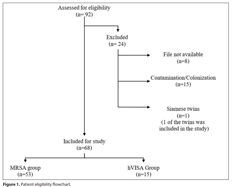
A total of 92 patients with culture-confirmed MRSA infection were identified and 68 of these included the study. Re-evaluation of retained isolates using both screening techniques and reference PAP-AUC revealed that 15 isolates (22%) were hVISA, while 53 isolates were solely MRSA. There were no VISA isolates (Figure 1).
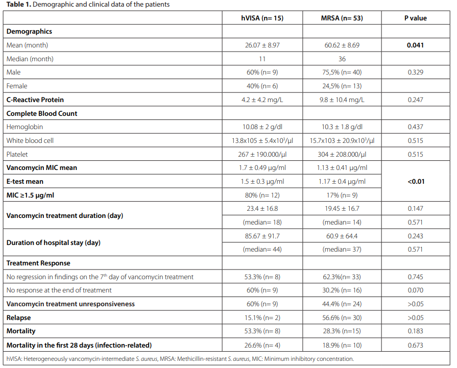
Mean age of patients with hVISA was younger than that of those with MRSA; their mean ages were 26.07 ± 8.97 and 60.62 ± 8.69 months, respectively. There were eight newborn patients in the MRSA group and two in the hVISA group (Table 1).
Mean MICs obtained by liquid microdilution method were 1.13 ± 0.41 micrograms/milliliter in children with MRSA infection and 1.70 ± 0.49 micrograms/milliliter in those with hVISA. Gradient diffusion testing yielded similar patterns, with values of 1.17 ± 0.40 and 1.50 ± 0.30 micrograms per milliliter, respectively. MIC values determined by gradient diffusion were significantly higher in children with hVISA (p< 0.01), supporting the association between elevated values and the presence of subpopulations with diminished responsiveness to vancomycin (Table 1).
Vancomycin was administered to infants and children at a dose of 60-70 mg/kg/day, given intravenously four times a day in divided doses, as recommended by the American Academy of Pediatrics. For neonates, it was given according to the appropriate week following a loading dose. Day-seven treatment failure occurred in 62.3% of the children with hVISA and in 53.3% of the MRSA group. By the end of therapy, treatment failure was documented in 60% of the hVISA group compared with 30.2% of the MRSA group. One patient was changed to linezolid treatment in the MRSA group, while no patients in the hVISA group were changed to this treatment. Treatment was unsuccessful in nine patients in hVISA group and 15 patients in MRSA group, and recurrent bacterial growth was observed. Duration of hospitalization was longer in the hVISA group compared with the MRSA group. Mortality rate within 28 days was 53.3% and 28.3% in hVISA and MRSA groups, respectively (Table 1).
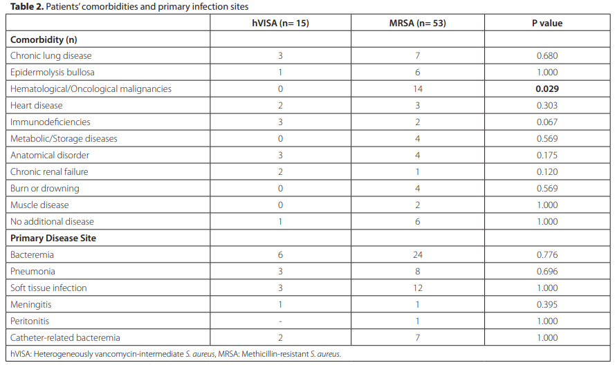
Although underlying medical conditions were generally similar between the groups, hVISA was more common in males (60%). Clinical manifestations tended to be more severe in children with hVISA, who showed higher frequencies of bloodstream infection, pneumonia, and soft-tissue infection. Baseline laboratory findings—including CRP, white blood cell count, hemoglobin concentration, and platelet count—were comparable between the two groups (Table 2)
In 88.2% of the cases, antimicrobial treatment was initiated within the first hour of the diagnosis. Mortality rate was 37.5% in patients who did not receive treatment within the first hour.
In our study, the most significant differences between the hVISA and MRSA groups were hospitalization frequency (p= 0.022), history of vancomycin treatment within the last three months (p= 0.95), and admission to the intensive care unit (p= 0.066).
DISCUSSION
Decreased susceptibility to vancomycin in S. aureus has transformed the clinical management of methicillin-resistant infections and raised significant concerns about treatment efficacy. While this phenomenon has been extensively described in adults, pediatric data are limited, and its clinical implications in children are not well understood due to developmental differences in immune function, antimicrobial pharmacokinetics, and underlying comorbidities. In this context, the present study offers one of the comprehensive assessments of heterogeneously decreased susceptibility to vancomycin in children with culture-confirmed MRSA infection.
In our study, vancomycin susceptibility was evaluated using both screening methods and the PAP-AUC technique, which is widely regarded as the most sensitive method for detecting heteroresistant subpopulations. Although labor-in-
tensive and costly, this technique is essential for accurately identifying strains that may contribute to therapeutic failure. By applying this approach to stored isolates from pediatric patients, the study provides valuable insight into the prevalence and clinical implications of hVISA in childhood, an area where published data remain scarce.
A limited number of pediatric studies have assessed the presence of hVISA, and most have relied on screening tests alone. Jimenez-Truque et al. reported a prevalence of 2.82% using gradient diffusion, while Welsh et al. identified only one case among 22 children with bloodstream infection (10,11). Mirza et al. conducted the only pediatric study using the population analysis profile method and demonstrated that heteroresistance was strongly associated with higher vancomycin MIC values, reaching 53.6% among isolates with a value of 2 µg/mL (9). In the present study, a similar relationship was observed; hVISA exhibited significantly higher vancomycin MIC values by gradient diffusion (p< 0.01), supporting the association between elevated values and reduced susceptibility.
In our study, mean age was significantly lower in the hVISA group compared with the MRSA group (26.07 ± 8.97 months vs. 60.62 ± 8.69 months, p= 0.041), indicating a notable age imbalance between the study groups. This difference represents a potential confounding factor that should be considered when interpreting the clinical outcomes. Age was not adjusted for in the primary analysis; therefore, part of the observed differences in treatment response, hospitalization frequency, and mortality may be influenced by age-related factors rather than pathogen-related characteristics alone. Younger patients, particularly infants and toddlers, have immature immune responses, limited physiological reserves, and a higher susceptibility to severe infections, which may predispose them to poorer outcomes independent of antimicrobial resistance patterns. In addition, the inclusion of newborns and very young children—who constitute a clinically distinct and more vulnerable subgroup—may have contributed both to outcome severity and to the lack of statistical significance in some comparisons.
Although treatment failure did not differ statistically between the groups, failure rate was clinically higher in hVISA infections (60% vs. 30.2%). This finding is consistent with adult studies, such as those by Casapao et al., which reported vancomycin treatment failure in more than 80% of heteroresistant bloodstream infections (12). Despite statistically not significant, high mortality rates were observed in the hVISA group (53.3%). This statistical insignificance in comparison may be attributed to the small number of hVISA cases and the inclusion of newborns, who represent a clinically distinct and physiologically vulnerable subgroup.
An important observation was the higher frequency of heteroresistance in children under one year of age. To date, no study has specifically examined the prevalence of heteroresistant strains in newborns, and the present findings highlight the need for further research in this population. Age-related physiological differences, including immature immune function and altered pharmacokinetics, may influence both the host response and the selection of resistant bacterial subpopulations. This underscores the limitations of applying adult-derived susceptibility breakpoints and treatment thresholds to newborns and infants.
Fatsis-Kavalopoulos et al. investigated the prevalence, risk factors, and clinical effects of the hVISA phenotype in a study involving 842 adult patients with MRSA bacteremia (13). This prospective study revealed strong positive correlations between previous MRSA treatment, vancomycin exposure, and especially vancomycin MIC. Patients in the hVISA group had higher mortality rates and similarly longer durations of bacteremia. Similar to our study, patients with MRSA-diagnosed antibiogram results were re-evaluated using the PAP-AUC method to assess their clinical status. The number of hVISA cases in this study was 184. Compared to our study, this is a significant clinical study due to its larger patient count and prospective nature. Our study, however, is the first in the literature to examine the pediatric and especially the neonatal period.
CONCLUSION
The findings of this study reinforce the importance of recognizing hVISA as a clinically significant phenomenon in pediatric practice. High vancomycin MIC values and inadequate clinical response should lead clinicians to consider the possibility of hVISA infection and evaluate alternative treatment strategies. This study provides the first pediatric data on the clinical significance of hVISA in Turkiye. Multicenter studies with larger pediatric cohorts may give better clinical outcomes associated with hVISA in the childhood.