
 TR
 EN


ABSTRACT
Objective
Community-acquired pneumonia is a major cause of morbidity in children, with Mycoplasma pneumoniae responsible for a substantial proportion of cases in school-aged children. Although most infections are mild, extrapulmonary complications, including cold agglutinin–mediated hemolysis, may occur. This study aimed to describe pediatric cases of M. pneumoniae pneumonia complicated by cold agglutinin disease and highlight diagnostic considerations.
Materials and Methods
This case series included four pediatric patients hospitalized with clinical and radiological findings consistent with M. pneumoniae infection. Dignosis was confirmed by polymerase chain reaction on nasopharyngeal swab samples. Complete blood counts were obtained at admission and during follow-up, and hemoglobin/ hematocrit (Hb/Hct) mismatches prompted evaluation for cold agglutinin disease. Laboratory assessments included peripheral blood smears, lactate dehydrogenase, indirect bilirubin, and direct Coombs testing. Chest radiographs were reviewed, and demographic, clinical, laboratory, treatment, and outcome data were recorded.
Results
All patients exhibited Hb/Hct mismatch during hospitalization, and direct Coombs testing was positive in three cases. Hematocrit levels increased markedly when blood samples were analyzed using warmed tubes, supporting the presence of cold agglutinin–mediated hemolysis. Peripheral blood cell lines other than erythrocytes were within normal limits. No patient required transfusion or specific therapy for hemolysis; standard antimicrobial treatment and supportive measures, including avoidance of cold exposure, were sufficient.
Conclusion
Cold agglutinin disease is a rare but clinically relevant extrapulmonary complication of M. pneumoniae infection in children. Recognition of Hb/Hct mismatch and confirmation using warmed sample analysis can facilitate accurate diagnosis and prevent unnecessary interventions. Awareness of this entity is essential for timely identification, appropriate management, and avoidance of overtreatment in pediatric pneumonia cases.
KEYWORDS
Anemia, autoimmune hemolytic, Mycoplasma pneumoniae, cold agglutinin disease, red blood cells
INTRODUCTION
Community-acquired pneumonia is among the leading causes of morbidity and mortality in childhood. While Streptococcus pneumoniae, Haemophilus influenzae, and Staphylococcus aureus are among the predominant bacterial pathogens in pediatric pneumonia, Mycoplasma pneumoniae is responsible for nearly 40% of cases in children older than five years of age (1). Although most lower respiratory tract infections caused by M. pneumoniae are mild, in some cases, they may progress to severe clinical conditions, including acute respiratory distress syndrome. On the other hand, extrapulmonary complications may occur in about 25% of the patients (1). Cold agglutinin hemolysis is one of these complications and typically presents as mild and transient hemolysis, although in rare cases it may follow a severe course (2).
Cold agglutinin disease is a form of autoimmune hemolytic anemia characterized by the production of antibodies directed against erythrocyte surface antigens. The phenomenon of blood agglutination at low temperatures was first described in the early 20th century, and in 1950, it was shown that cold agglutinins may develop in patients with atypical pneumonia. In addition, viral and bacterial infections, as well as certain lymphomas, have been implicated in the development of cold agglutinin disease (3).
Hemolysis secondary to M. pneumoniae infection is mediated by cold agglutinin antibodies targeting the I antigen on erythrocyte surfaces. The I antigen, a carbohydrate moiety expressed on both erythrocytes and respiratory epithelial cells, also functions as a receptor facilitating mycoplasma adherence. In this setting, M. pneumoniae may act as an adjuvant, inducing an antibody response against the I antigen (4).
This case series describes four patients hospitalized and treated for M. pneumoniae infection who exhibited a mismatch between hemoglobin (Hb) and hematocrit (Hct) values on complete blood count (CBC). By presenting their clinical courses and laboratory findings, this series aims to increase awareness of secondary cold agglutinin disease in patients with Hb/Hct mismatches and a recent history of lower respiratory tract infection, thereby helping to prevent unnecessary diagnostic and therapeutic interventions.
MATERIALS AND METHODS
This case series included four pediatric patients who were admitted to our hospital with clinical and radiological findings consistent with M. pneumoniae infection and were found to have cold agglutinin hemolysis. The diagnosis of M. pneumoniae infection was confirmed by polymerase chain reaction (PCR) analysis performed on nasopharyngeal swab samples in addition to compatible clinical presentation.
CBC were obtained for all patients at the time of admission and during follow-up. A mismatch between hemoglobin (Hb) and hematocrit (Hct) values was noted, prompting further evaluation for cold agglutinin disease. Peripheral blood smear examinations, lactate dehydrogenase (LDH), and indirect bilirubin levels were assessed to support the diagnosis of hemolysis. The direct Coombs test was also performed.
Chest radiographs were evaluated to document pulmonary involvement. Demographic characteristics, clinical presentations, laboratory findings, treatment approaches, and outcomes of all cases were recorded and analyzed descriptively.
For the research, ethics committee approval with protocol number 2025/19/1 was received from the non-interventional clinical research ethics committee of Samsun University Non-Invasive Clinical Research on 01.10.2025.
Case 1
A 13-year-old female patient presented to our outpatient clinic with intermittent fever and cough ongoing for 20 days. She had previously visited external centers twice and had received outpatient antibiotic therapy. She had no known chronic diseases. On physical examination, her temperature was 38.2 °C, and bilateral fine crackles were detected. Initial CBC showed: Hb 11.7 g/Dl, red blood cell (RBC) 3.18 million/ µL, Hct 26.2%, mean corpuscular volume (MCV) 82.4 fL, white blood cell (WBC) 10.9 ×10³/µL, platelet (PLT) 252 ×10³/µL, reticulocyte 2.52%. Additional laboratory findings were: urea 25.2 mg/dL, creatinine 0.75 mg/dL, alanine aminotransferase (ALT) 28 U/L, aspartate aminotransferase (AST) 41 U/L, C-reactive protein (CRP) 63 mg/L, and erythrocyte sedimentation rate (ESR) 75 mm/h. Posteroanterior (PA) chest radiograph revealed
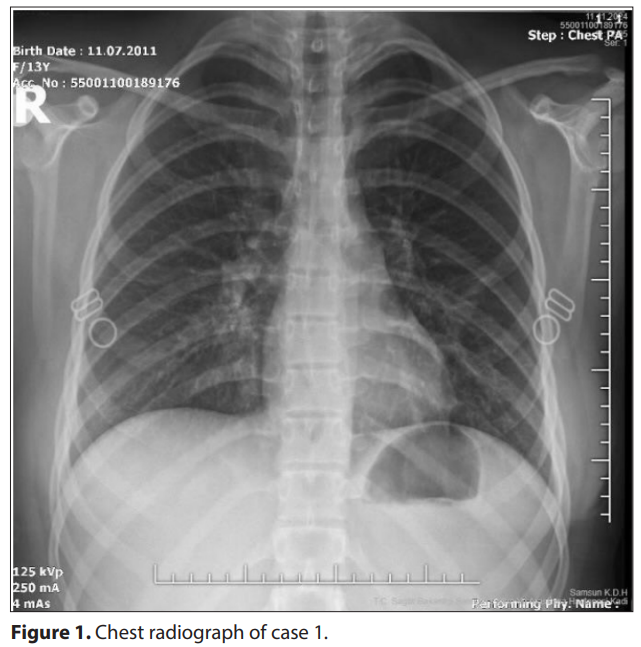
infiltration in the lower zone of the right lung, in the paracardiac region (Figure 1). The patient was hospitalized with the diagnosis of community-acquired pneumonia.
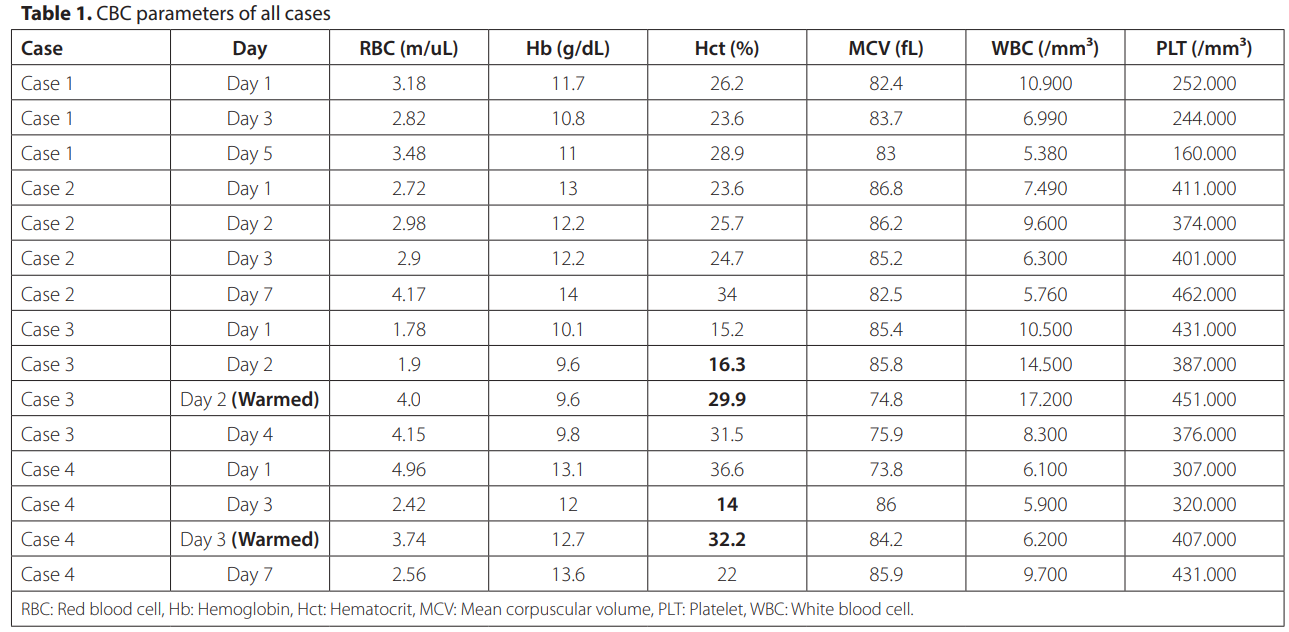
Ceftriaxone and oral clarithromycin treatment were initiated. Due to the mismatch between hemoglobin and hematocrit values, further tests were performed: total bilirubin 0.8 mg/dL, indirect bilirubin 0.66 mg/dL, LDH 490 U/L, and direct Coombs test was positive. Nasopharyngeal swab obtained at admission was positive for M. pneumoniae. Peripheral blood smear performed for hemolysis was evaluated as normal. The patient’s hemogram parameters at admission and during follow-up are presented in Table 1.
Case 2
A 10-year-old boy presented with a 10-day history of cough and loss of appetite. Four days earlier, he had been evaluated at another facility and prescribed oral antibiotics for acute upper respiratory tract infection. As his symptoms persisted, he was admitted to our emergency department. Examination revealed diminished breath sounds and crepitant rales over the left lung. On admission, his general condition was poor, oxygen saturation was 97%, and he was in no respiratory distress. Ceftriaxone, clindamycin, and oral clarithromycin were initiated. Laboratory investigations revealed: Hb 13 g/dL, RBC 2.72 million/µL, Hct 23.6%, MCV 86.8 fL, WBC 7.49 ×10³/µL, PLT 411 ×10³/µL.
Renal and hepatic function tests were unremarkable. Inflammatory markers included CRP 40 mg/L and ESR 80 mm/h. A PA chest radiograph demonstrated consolidation consistent with pneumonic infiltration in the lower zone of the left lung. The patient was hospitalized with the diagnosis of community-acquired pneumonia.
Because of an Hb/Hct mismatch, further testing was performed, which showed total bilirubin 0.37 mg/dL, indirect bilirubin 0.31 mg/dL, LDH 190 U/L, and a negative direct Coombs test. PCR of a nasopharyngeal swab confirmed M. pneumoniae. The patient’s hemogram parameters at admission are summarized in Table 1.
Case 3
A 17-year-old female patient presented with cough, fever, loss of appetite, and fatigue for 10 days. She had visited an external center one week earlier and had been prescribed oral antibiotics; however, since her symptoms did not improve, she was admitted to our hospital. On examination, widespread crepitant rales were detected. Her oxygen saturation was 98%, and she had no respiratory distress. Laboratory tests at admission showed: Hb 10.1 g/dL, RBC 1.78 million/µL, Hct 15.2%, MCV 85.4 fL. Renal and liver function tests were unremarkable. CRP was 93 mg/L and ESR 100 mm/h. PA chest radiograph revealed an image consistent with infiltration in the lower–middle zone of the right lung. She was hospitalized with the diagnosis of pneumonia.
Ceftriaxone, clindamycin, and oral clarithromycin were started. The initial direct Coombs test was negative. Nasopharyngeal swab collected at admission tested positive for M. pneumoniae. On the second day of hospitalization, repeat direct Coombs test became positive.
Because the mismatch between hemoglobin and hematocrit values was thought to be related to cold agglutinins, the laboratory was contacted for warmed sample analysis. While the patient’s Hct was initially 16.3%, repeat CBC performed on a blood sample incubated at 37°C showed approximately a twofold increase in RBC count and Hct rising to 29.9%. These findings supported the presence of cold agglutinins. The patient was advised to wear warm clothing, and her current treatment was continued.
The patient’s hemogram parameters at admission and during follow-up are summarized in Table 1.
Case 4
A 10-year-old male patient presented to the emergency department with a one-week history of fever and cough. On physical examination, there were fine crackles. Initial laboratory evaluation revealed: Hb 13.1 g/dL, RBC 4.96 million/µL, Hct 36.6%, MCV 73.8 fL, WBC 6.1 ×10³/µL, PLT 307 ×10³/µL, urea 17.2 mg/dL, creatinine 0.48 mg/dL, ALT 15.1 U/L, AST 28.1 U/L, and CRP 40 mg/L. PA chest radiograph obtained in the emergency department. The patient was admitted with a presumptive diagnosis of community-acquired pneumonia. Empirical therapy with ceftriaxone and oral clarithromycin was initiated.
On the third day of hospitalization, follow-up tests revealed a mismatch between hemoglobin and hematocrit values (Table 1). Direct Coombs testing performed to evaluate hemolysis was positive. Considering that the hemoglobin/hematocrit discrepancy could be due to cold agglutinin, a repeat complete blood count was performed using a warmed blood sample. While the initial Hct was 14%, analysis of the warmed sample revealed an Hct of 32.2%, supporting the diagnosis of cold agglutinin disease. On the fourth day of hospitalization, nasopharyngeal swab testing confirmed M. pneumoniae.
DISCUSSION
In our series, four patients hospitalized with pneumonia who exhibited hemoglobin/hematocrit mismatch were found to have M. pneumoniae detected in respiratory swab samples. In all cases, hematocrit levels improved during pneumonia treatment without the need for additional therapy targeting cold agglutinin disease. Although cold agglutinin antibody testing could not be performed in our center, the diagnosis was supported by a marked increase in hematocrit values when complete blood counts were repeated using blood samples incubated at nearly 37 °C.
M. pneumoniae typically causes upper or lower respiratory tract infections in school-aged children over five years of age (1). The bacterium adheres to the host respiratory epithelium, inducing direct cell membrane damage through oxidative stress and contributes to the development of bronchopneumonia and pneumonia via inflammation mediated by alveolar macrophages. Complications affecting various systems, including the central nervous system, gastrointestinal tract, and hematologic system, have also been reported. The pathophysiology of extrapulmonary manifestations involves indirect immune-mediated injury, vascular occlusion due to vasculitis or thrombosis, and locally induced inflammatory cytokines. M. pneumoniae can trigger local vasculitis or thrombosis through cytokines such as TNF-α and IL-8, without causing a systemic hypercoagulable state. As a result, abdominal pain, chest pain, neurological symptoms, and, very rarely, priapism have been reported (5).
M. pneumoniae is known to cause cold agglutinin disease as an antibody-mediated extrapulmonary complication. Cold agglutinin disease accounts for approximately 15% of autoimmune hemolytic anemias, with an estimated incidence of 1 in 1.000.000 individuals (6). Antibodies developed against M. pneumoniae react with I antigens on the erythrocyte surface, leading to hemolysis. In children, cold agglutinin disease can be triggered by infections such as M. pneumoniae, influenza virus, adenovirus, hepatitis B and C, HIV, EBV, and CMV (3). The four cases presented in our series were all associated with M. pneumoniae infection and were encountered within the past year. Epidemics of M. pneumoniae are known to occur every three–seven years. We hypothesize that the unusual occurrence of four cases of this rare complication is likely related to an increase in M. pneumoniae infections this year.
Hematologic complications of M. pneumoniae infection may, although rarely, include pancytopenia, thrombocytopenia, thrombocytosis, or leukemoid reactions (2,7,8). In our cases, however, non-erythroid cell lines remained within normal ranges. Cold agglutinin–mediated hemolytic anemias generally tend to resolve spontaneously, but patients with severe anemia may require transfusion (9). Blood transfusion should be administered cautiously in such cases, as it can increase the risk of hemolysis. Intravenous immunoglobulin and plasmapheresis have been used in life-threatening, transfusion-resistant cases, resulting in an increase in hematocrit (10). A normal hemoglobin is generally expected to be 1/3 of the Hct (11). Additionally, cases in the literature have demonstrated that hematocrit values may increase from 7% to 30% when complete blood counts are repeated using warmed samples (12). In our series, two patients’ samples were reassessed using the water bath method, resulting in hematocrit increases from 16.3% to 29.9% and from 14% to 32.2%, respectively. This approach allows for a more accurate evaluation of hematocrit levels and may reduce unnecessary transfusion decisions. Although its efficacy has not been formally evaluated, patients are also advised to avoid cold exposure and wear warm clothing (6).
CONCLUSION
Cold agglutinin disease is a rare but important extrapulmonary complication of M. pneumoniae infection in children. Recognition of hemoglobin/hematocrit mismatch, together with supportive laboratory assessments such as warmed sample analysis, can aid in accurate diagnosis and prevent unnecessary interventions. One of the limitations of our study is that cold agglutinin titers cannot be studied in our hospital. Our case series demonstrates that this complication is generally self-limiting and can be effectively managed with standard antimicrobial therapy and supportive measures, including avoidance of cold exposure. Awareness of this entity among clinicians is essential to ensure timely identification, appropriate management, and avoidance of overtreatment in pediatric patients with M. pneumoniae pneumonia.