
 TR
 EN


ABSTRACT
Objective
Sinusitis is a common condition in childhood that may lead to various serious complications. While orbital cellulitis is more frequently observed, subdural empyema, though rare, represents a life-threatening condition. We report the case of an eight-year-old girl who developed orbital cellulitis and subdural empyema secondary to pansinusitis. The patient initially presented with meningismus, fatigue, and rhinorrhea, later progressing to right eye swelling, headache, drowsiness, vomiting, and fever. Despite initial treatment for meningitis, her symptoms worsened, prompting referral to our center. Imaging studies confirmed pansinusitis complicated by subdural empyema. The patient required surgical drainage of both the empyema and the sinuses in addition to intravenous antibiotics. After six weeks of therapy, follow-up imaging demonstrated complete resolution, and she was discharged without sequelae. This case highlights the severity of complications of pansinusitis in children and underscores the importance of early recognition, timely imaging, and a multidisciplinary approach for optimal outcomes.
KEYWORDS
Keywords: Orbital cellulitis, paranasal sinus diseases, subdural empyemae
INTRODUCTION
Sinusitis is a prevalent condition that may present with a variety of symptoms, including facial pressure, nasal congestion, and purulent discharge. Most cases resolve without significant complications; however, a subset of individuals may experience severe consequences. Among the most concerning complications of pansinusitis are orbital complications. These are orbital cellulitis, preseptal/periorbital cellulitis, subperiosteal abscess, and optic neuritis (1).
Subdural empyema arises from the accumulation of pus between the dura mater and the arachnoid mater. Although the widespread availability of potent antibiotics has significantly decreased the occurrence of these complications, they still pose a potentially life-threatening risk (2). Herein, we present an 8-year-old girl who developed orbital cellulitis, subdural empyema, and pneumocephalus as secondary complications arising from pansinusitis.
CASE REPORT
A previously healthy eight-year-old child presented to our hospital exhibiting signs of meningismus. The patient reported a runny nose and fatigue that had persisted for four days. Two days prior to admission, she developed right eye swelling, which was subsequently followed by headache, drowsiness, vomiting, and fever. Initially diagnosed with meningitis at the referring center, the patient had received a single dose of ceftriaxone. However, due to the severity and progression of symptoms, she was referred to our center.
Upon admission, the patient was lethargic, and the Glasgow Coma Score was 12. Her vital signs revealed a temperature of 38.2 °C, blood pressure of 100/60 mmHg, and pulse rate of 110 beats per minute. Eye examination revealed edema, hyperemia, and increased temperature in her right eye. Oropharyngeal examination demonstrated significant postnasal discharge. Neurological examination revealed neck stiffness and positive Brudzinski and Kernig signs. Pupillary light reflexes were normal, and eye movements were intact. The patient’s personal and family medical histories were unremarkable.
Laboratory results were as follows: White blood cell count was 14.7 x 10³ /mm3 (normal range 4-10 x 103 /mm3 ); hemoglobin, 11.2 g/dL (normal range 11.5–15.5 g/dL); platelet count: 99 x 103 /mm3 (normal range 150 – 450 x 103 /mm3 ); C-reactive protein: 144 mg/L (normal range 0–5 mg/L); and procalcitonin: 151 µg/L (normal range 0–1 µg/L). Cranial computed tomography (CT) scan revealed linear loculations and air images between the right cerebral hemisphere and falx, suggestive of subdural empyema. Fundoscopic examination revealed no papilledema. A lumbar puncture was performed. Cerebrospinal fluid (CSF) pressure was normal, but the appearance was cloudy. Direct examination of CSF revealed a significant increase in neutrophils (165/mm³). CSF analysis revealed the following: A protein level of 38 mg/dL (normal range of 15–45 mg/dL) and a glucose level of 70 mg/dL (normal range of 50– 70% of serum glucose; the patient’s blood glucose level was 113 mg/dL). CSF culture yielded no microorganisms. Given the severity of the patient’s condition, empirical therapy with ceftriaxone, vancomycin, and metronidazole was initiated. In addition to antibiotics, dexamethasone was administered every six hours for four days.
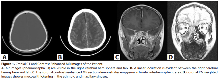
Magnetic resonance imaging (MRI) and CT scans were performed. Brain CT revealed air bubbles and linear fluid loculation in the right interhemispheric area (Figure 1A, B). Diffusion MRI showed restricted diffusion in the loculated fluid, indicating an infectious collection. Contrast-enhanced MRI demonstrated infectious loculation measuring 3.5 mm in depth and dural enhancement consistent with subdural empyema. Mucosal thickening in the sinuses was observed (Figure 1C, D).
By the third day after the initial presentation, the swelling in the right eye had worsened and was accompanied by fever. New imaging studies were performed to rule out additional complications. Contrast-enhanced brain CT scans revealed significant edema in the right preseptal region of
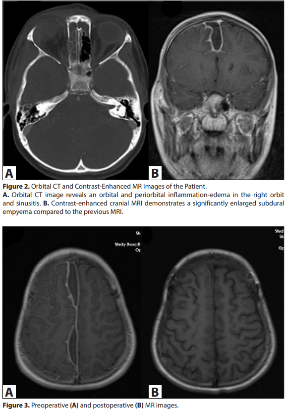
the orbit and a fluid collection measuring approximately 13 x 9 mm in the superior part of the right intraorbital extracanalicular space. The subdural empyema measured 9 mm in thickness. Subsequent contrast-enhanced cranial MRI demonstrated a significantly enlarged loculation consistent with subdural empyema as compared with the previous cranial MRI (Figure 2).
The patient’s subdural empyema was successfully treated with neurosurgical drainage and sinus drainage performed by the ear, nose, and throat department. A microbiological culture was obtained during the drainage of the subdural empyema, but no growth was detected. This finding was attributed to the antibiotic therapy administered from the outset. A radiological examination conducted 28 days after surgery showed complete resolution of the subdural empyema (Figure 3). The patient completed a six-week course of antibiotic therapy and was discharged from the hospital without any sequelae.
DISCUSSION
Subdural empyema is a serious condition characterized by an accumulation of pus between the dura mater and arachnoid mater. Around 35-65% of subdural empyema cases are attributed to sinusitis (3). Patients displaying symptoms such as altered eye movements, visual disturbances, proptosis, neck stiffness, headaches, and changes in consciousness should prompt consideration of various potential diagnoses, including preseptal cellulitis, orbital cellulitis, subperiosteal abscess, orbital abscess, and cavernous sinus thrombosis (4).
The causative organisms responsible for subdural empyema often vary depending on the patient’s age and the source of the infection. Paranasal sinuses commonly harbor a variety of bacteria, including alpha-hemolytic streptococci, anaerobic streptococci, non-hemolytic streptococci, Staphylococcus aureus, Bacteroides species, and Enterobacteriaceae. When subdural empyema is associated with otitis media, it is crucial to rule out alpha-hemolytic streptococci and Pseudomonas aeruginosa as potential culprits (5). In this particular case, ceftriaxone, vancomycin, and metronidazole were administered. In addition to antibiotics, dexamethasone was administered to reduce the risk of meningitis sequelae, such as hearing loss, seizures, and intellectual disability. A recent cohort study demonstrated that the administration of dexamethasone within 12 hours of initiating antibiotic therapy reduced mortality in pneumococcal meningitis (6). In our patient, given the unstable clinical condition, we anticipated potential benefit and administered dexamethasone despite prior antibiotic therapy. The recommended overall treatment duration typically ranges from six to eight weeks, with intravenous therapy administered for two to six weeks followed by a transition to oral antibiotics. The decision to switch from intravenous to oral antibiotics should be guided by several factors, including the patient’s neurological status, the absence of fever, and evidence from CT or MRI scans demonstrating resolution of the purulent collection. Serial imaging studies are essential for monitoring treatment progress and assessing the need for continued antibiotic therapy (5).
In patients presenting with potential orbital or intracranial complications, cranial CT scans should be performed for neurosurgical evaluation. Contrast-enhanced CT is particularly useful in such cases as it can effectively visualize the extent of inflammation, abscess formation, and any bony involvement. MRI offers superior tissue differentiation and can help distinguish between malignant masses and inflammatory conditions such as subdural empyema (4). In our patient, complications had developed, and since rapid intervention in such cases is critical, repeated imaging was required.
Even though certain cases of subdural empyema may respond favorably to antibiotic therapy alone, surgical drainage remains essential for orbital abscesses (7). Conservative or nonoperative management may be considered for patients who meet specific criteria, including overall good health, no significant neurological deficits, a small empyema collection, localized lesions except in the posterior fossa, and a favorable response to antibiotics (8). However, in our patient, there was no positive response to antibiotics, and the collections increased after three days, indicating a need for a different approach due to lack of improvement.
Our main message is that subdural empyema is a potentially life-threatening complication that demands prompt recognition and aggressive treatment. A comprehensive medical history, a thorough physical examination, and a high index of clinical suspicion are crucial for identifying subdural empyema. Imaging studies play a pivotal role in confirming the diagnosis and assessing the extent of the infection. Early diagnosis and treatment are important to minimizing the risk of longterm sequelae.