
 TR
 EN


INTRODUCTION
Dear Editor,
Varicella has been identified as a major risk factor for invasive Streptococcus pyogenes infections, with reported complications including cellulitis, necrotizing fasciitis, pyomyositis, pneumonia, empyema, endocarditis, and sepsis (1,2). Herein, we report a pediatric case of pyomyositis complicating scarlet fever superimposed on an active varicella infection.
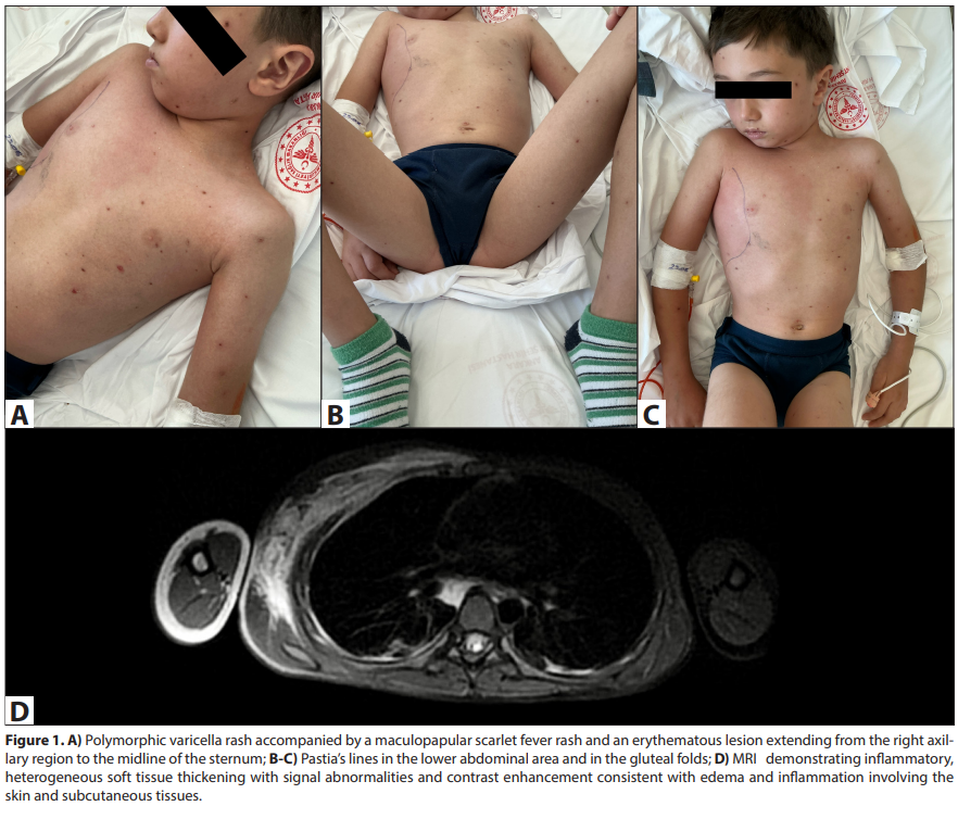
A six-year-old boy, previously vaccinated according to the national immunization schedule including a single dose of varicella vaccine, presented to an outside facility with a vesicular rash that began on the head and neck and subsequently spread over the entire body, leading to a diagnosis of varicella. His history revealed fever and rash for the past four days, with the onset of pain originating from the axillary region and radiating to the neck over the preceding two days. His sibling had experienced varicella two weeks earlier. The patient was referred to our hospital due to a progressive lesion on the neck and chest. On physical examination, there were vesicular lesions in various stages of crusting consistent with varicella. In addition, hyperemic maculopapular rashes that blanched on pressure were observed on the trunk and neck, as well as on the arms and legs, with a positive sandpaper-like texture (Figure 1A). Pastia’s lines were noted in the abdominal and inguinal folds (Figure 1B,C). An erythematous skin lesion extending from the right axillary region toward the midline of the sternum was present, accompanied by localized warmth, swelling, and marked tenderness on palpation. The patient experienced severe pain on the right shoulder and neck movements, rendering him unable to lift his arm or turn his neck. S. pyogenes was detected in a respiratory PCR test performed on an oropharyngeal swab sample. The patient was diagnosed with concurrent varicella and scarlet fever complicated by secondary cellulitis and pyomyositis, and treatment with ceftriaxone, clindamycin, and acyclovir was initiated.
Laboratory tests revealed the following: leukocytes 23.94 × 10³/µL, neutrophils 20.79 × 10³/µL, lymphocytes 2.05 × 10³/ µL, hemoglobin 12.8 g/dL, platelets 226.000/µL, and C-reactive protein 207 mg/L. Follow-up examinations performed on the same day showed that the painful, erythematous cellulitic lesion had markedly progressed within hours, extending beyond the pectoral region toward the midline of the sternum and further inferiorly. The patient was transferred to the intensive care unit. No pathology was detected on ultrasonography of the right shoulder joint. Neck and thoracic magnetic resonance imaging (MRI) revealed inflammatory, heterogeneous soft tissue thickening measuring up to 15 mm in thickness, with signal intensities and contrast enhancement consistent with edema and inflammation. These findings extended from both inferior cervical regions, the right axillary and anterior axillary areas, and the anterior, lateral, and posterior walls of the right chest, inferiorly to the lateral and posterior abdominal wall, and posteriorly to the dorsal region. The process involved the skin and subcutaneous tissues, as well as intermuscular planes between the chest and abdominal wall muscles, predominantly affecting the pectoralis, intercostal, and latissimus dorsi muscles (Figure 1D). The MRI findings supported the diagnosis of pyomyositis. Blood cultures remained sterile. As the varicella lesions crusted, acyclovir was discontinued on day seven. During follow-up, the patient’s joint pain and skin lesions resolved completely.
Secondary bacterial infections in varicella are most caused by S. pyogenes and Staphylococcus aureus. While there are numerous reports describing cutaneous complications due to S. pyogenes infection, cases of concurrent varicella and scarlet fever have been rarely documented (3-5). Reported complications of concurrent varicella and scarlet fever include empyema, cellulitis, severe swelling of the inguinoscrotal region, and widespread monomorphic vesicular eruptions superimposed on the erythema of scarlet fever (3-5). Streptococcal pyomyositis, necrotizing pyomyositis, and necrotizing fasciitis
are among the rare but severe complications of varicella, and their management may require fasciotomy, abscess drainage, and hyperbaric oxygen therapy (2). In the present case, we aimed to highlight that varicella and scarlet fever can, although rarely, occur simultaneously and may be complicated by pyomyositis. In countries such as Türkiye, where only a single dose of varicella vaccine is administered, the introduction of a routine second dose could contribute to the prevention of breakthrough varicella infections and associated complications.