
 TR
 EN


INTRODUCTION
A 10-year-old boy presented to the rheumatology outpatient clinic with complaints of inability to bear weight on his feet, swelling of the ankles, fever, and fatigue. His history revealed intermittent low back and back pain, fever reaching 38°C, and weight loss for approximately three months. He had no history of recurrent infections, trauma, or connective tissue disease.
Physical examination revealed swelling and limited range of motion in the ankles, as well as tenderness in the sacroiliac region. Laboratory investigations showed an elevated white blood cell count (11.900/mm³) with lymphocytic predominance. C-reactive protein (82.2 mg/L) and erythrocyte sedimentation rate (59 mm/hour) were elevated. Hemoglobin (8.7 g/dL) and hematocrit (29%) levels were below normal. Transthoracic and transesophageal echocardiography demonstrated extensive vegetations on the aortic valve extending into the ascending aorta, together with aneurysmal dilatations. Blood cultures were positive.
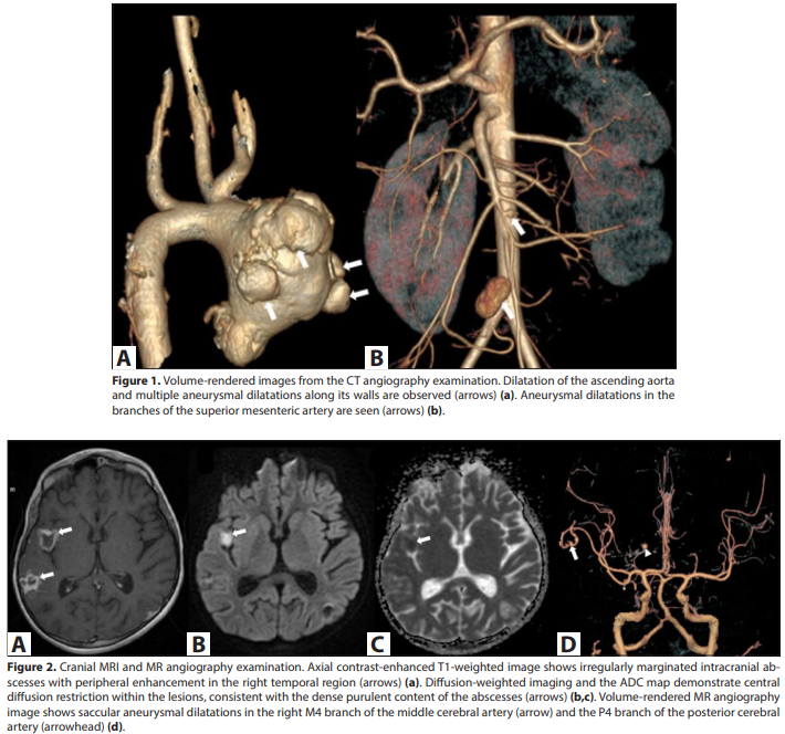
On the fifth day of antibiotic therapy, the patient developed severe abdominal pain, nausea, and vomiting. An emergency abdominal ultrasonography (US) examination revealed no sonographic findings suggestive of causes of acute abdomen, such as acute appendicitis, ileus, or an intra-abdominal abscess. Due to persistent symptoms, abdominal computed tomography (CT) and CT angiography were performed, revealing aneurysmal dilatations of the ascending aorta and an approximately 18×13 mm partially thrombosed saccular aneurysmal dilatation associated with the distal segment of the superior mesenteric artery. The contrast-filled patent lumen measured approximately 16×12 mm. Approximately 30 mm cranial to this aneurysm, a second lobulated saccular aneurysmal dilatation measuring 4×3 mm and associated with the superior mesenteric artery was also detected (Image 1).
On the tenth day of hospitalization, the patient underwent cardiac surgery. The destroyed aortic valve and infected tissues in the ascending aorta were excised, and mechanical aortic valve replacement was performed simultaneously with supracoronary ascending aortic graft replacement.
Five days after cardiac surgery, the patient developed altered mental status and seizures. Contrast-enhanced cranial MRI demonstrated irregularly marginated, peripherally enhancing abscesses with central diffusion restriction, located in the cortical and subcortical regions of both cerebral hemispheres; the largest measured approximately 15 mm and was located in the right temporal lobe. Magnetic resonance imaging (MR) angiography revealed millimetric saccular aneurysms in the M4 segment of the right internal carotid artery and the P4 segment of the posterior cerebral artery (Image 2).
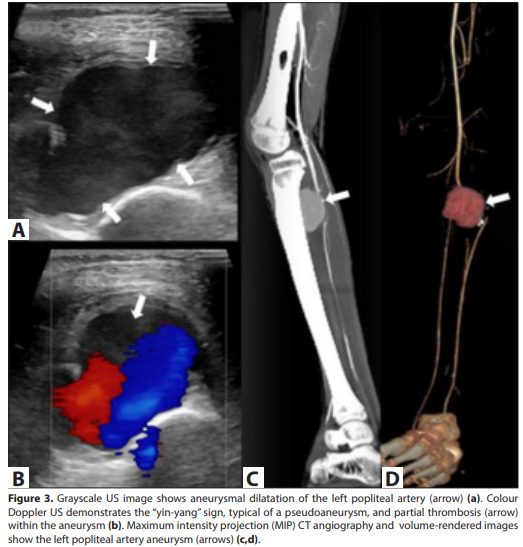
During follow-up, blood cultures became negative and inflammatory markers decreased. Ten days after cardiac surgery, the patient developed severe left leg pain. Emergency colour Doppler US revealed a wide-necked pseudoaneurysm
measuring approximately 55×32 mm, associated with the mid-to-distal segment of the left popliteal artery, without a clearly discernible wall. A thrombosed area was observed at the periphery of the lesion, and a characteristic “yin-yang” (bidirectional swirling flow) pattern was seen within its lumen. Lower-extremity CT angiography demonstrated a 42×30 mm saccular aneurysmal dilatation in the left popliteal fossa associated with the popliteal artery (Image 3).
Considering the patient’s history, physical examination, and radiological findings, what is your diagnosis?
DIAGNOSIS: Mycotic aneurysms secondary to infective endocarditis
Short discussion
A mycotic aneurysm is an infectious aneurysm that develops as a result of microorganisms colonising the arterial wall. Although the term “mycotic” is used, the causative organisms are generally not fungi but bacterial species of *Staphylococcus* or *Streptococcus*. In infective endocarditis, septic emboli originating from valvular vegetations may reach the arterial wall, disrupting its integrity and frequently resulting
in pseudoaneurysm formation. Infected aneurysms may occur in the aorta, as well as in peripheral, cerebral, and visceral arteries (1).
Although infective endocarditis is rare in children, it can lead to life-threatening complications such as heart failure, systemic embolisation, abscesses, and mycotic aneurysms. Mycotic aneurysms require early diagnosis because of the risks of rapid growth, thrombosis, distal embolisation, and rupture. In particular, the new onset of abdominal or extremity pain in a patient with infective endocarditis should raise suspicion of septic embolisation and mycotic aneurysm (2). In our case, abdominal and left leg pain developing at different times constituted the clinical manifestations of multiple mycotic aneurysms involving the superior mesenteric and popliteal arteries.
Imaging plays a fundamental role in detecting aneurysms, determining their anatomical extent, and planning treatment. Colour Doppler US is a readily accessible and effective method, particularly for evaluating peripheral arteries. The bidirectional “yin-yang” flow pattern within the pseudoaneurysm lumen and the characteristic flow changes at the neck support the diagnosis. CT angiography enables evaluation of the aneurysm’s size, its relationship with the artery, mural thrombus, distal circulation, and other accompanying vascular foci with higher spatial resolution. On CT, infected aneurysms frequently appear as saccular or lobulated, irregular-walled, partially thrombosed lesions (3).
Mycotic aneurysms of the superior mesenteric artery are extremely rare and represent a recognised visceral complication of infective endocarditis. They may present with nonspecific symptoms such as abdominal pain, nausea, and vomiting; if rupture occurs, they carry a risk of intra-abdominal haemorrhage and high mortality (4).
Mycotic aneurysms of the popliteal artery are also rare and may present with extremity pain, swelling, distal ischaemia, or loss of pulse. In our case, the detection of monophasic flow in the arteries distal to the aneurysm and markedly reduced flow at the ankle level on Doppler US indicated compromised distal circulation.
Appropriate long-term antibiotic therapy is essential in the treatment of mycotic aneurysms. However, surgical or endovascular treatment may be performed after considering the aneurysm’s location and size, whether it causes clinical symptoms, its enlargement during follow-up, impairment of distal circulation, and the risk of rupture. Some small mycotic aneurysms may regress or thrombose with antibiotic therapy; however, standard criteria for determining which lesions can be safely managed with medical treatment alone are limited. Furthermore, negative blood cultures and decreasing inflammatory markers do not definitively indicate that existing aneurysms will regress or that new aneurysms will not develop. In the presence of uncontrolled infection or ongoing embolic complications, surgical elimination of the persistent infectious focus together with the vegetations may be required (2,4).
In our case, the aneurysms involving the superior mesenteric artery and its distal branches were placed under close imaging surveillance; during follow-up, they were observed to decrease in size and undergo thrombosis. In contrast, surgical treatment was performed for the pseudoaneurysm of the left popliteal artery because it was symptomatic and compromised distal arterial circulation. These findings demonstrate that mycotic aneurysms in the same patient may follow different clinical courses depending on their location and clinical effects, and that treatment should be individualised for each lesion.
This case is noteworthy because it demonstrates that septic embolisation secondary to infective endocarditis in childhood may cause multiple mycotic aneurysms involving both visceral and peripheral arteries in the same patient. Since improvement in clinical and laboratory findings does not completely exclude vascular complications, new abdominal or extremity pain developing during follow-up should be carefully evaluated. When clinically suspected, targeted colour Doppler US should be performed for peripheral lesions, while CT angiography should be used to assess the extent of the lesions and changes during treatment. Follow-up CT angiography should be considered if symptoms recur or progression of existing aneurysms is suspected.