
 TR
 EN


INTRODUCTION
A two-year-seven-month-old male patient presented with a progressively growing swelling in front of the right ear that had been present for approximately five months. Despite receiving oral amoxicillin-clavulanate treatments at different times, the lesion had not completely resolved. There was no known tuberculosis exposure or raw milk consumption, but his family was involved in livestock farming. Physical examination revealed a mass approximately 3 x 2 cm in size, with erythema and fluctuation, located in front of the right tragus. Other system examinations were normal. Ultrasonography and contrast-enhanced magnetic resonance imaging revealed a complicated cystic lesion in the right preauricular subcutaneous tissue, extending to the adjacent parotid gland and exhibiting predominantly peripheral contrast enhancement. The findings were primarily considered indicative of an infected type 1 branchial cleft cyst or preauricular sinus. The lesion was surgically excised. During the operation, a caseified lesion was observed that infiltrated the superficial parotid lobe, lacked a capsule, and had irregular borders. Histopathological examination revealed granulomatous inflammation with localized necrosis and giant cells. Tuberculosis polymerase chain reaction (PCR) test on abscess drainage material was positive, and Mycobacterium bovis grew in cultures. The patient, who had received the BCG vaccine, had a tuberculin skin test of 13 mm and a positive QuantiFERON
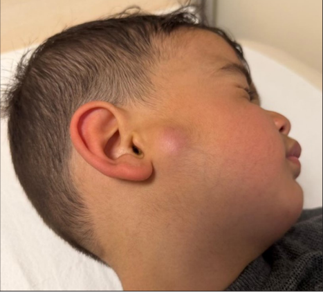
test. Thoracic computed tomography showed no pulmonary or mediastinal involvement. Acid-fast bacilli smear, tuberculosis PCR, and culture tests on three consecutive gastric fluid samples were negative. The patient, who was initiated on antituberculosis treatment, showed clinical improvement during follow-up, and no recurrence of the lesion was observed.
Preauricular and intraparotid tuberculosis is rare in children and can mimic infected branchial cleft cysts, preauricular sinuses, bacterial abscesses, or neoplastic masses. The absence of known tuberculosis contact and pulmonary involvement does not rule out the diagnosis. Mycobacterial infections should be considered in the differential diagnosis of chronic preauricular masses that do not respond to standard antibiotic treatment. Since the acid-fast bacilli smear, PCR, culture, and histopathology results can vary between samples in paucibacillar lesions, obtaining multiple aspirates and tissue samples from different areas of the lesion can improve diagnostic accuracy. Species identification is crucial for determining the appropriate treatment regimen and duration.